Kyle Wellmerling , Christian Lehmann , Ankur Singh , Brian J. Kirby
{"title":"用于从治疗性人类干细胞中无标记去除畸胎瘤形成细胞的微流控芯片","authors":"Kyle Wellmerling , Christian Lehmann , Ankur Singh , Brian J. Kirby","doi":"10.1016/j.regen.2020.100030","DOIUrl":null,"url":null,"abstract":"<div><p>Teratoma<span><span> formation remains a safety concern in therapeutic cells derived from human-induced pluripotent stem cells (hiPSCs). Residual Teratoma forming cells are present in small numbers in differentiated hiPSC cultures and yet are of significant roadblock to the manufacturing and clinical translation of stem cell therapies. Rare cells are often difficult to remove with standard flow cytometry or magnetic bead sorting techniques. Here, we first characterized time-dependent expression of a teratoma marker, stage-specific embryonic antigen (SSEA)-5, which binds the H type-1 </span>glycan<span> during neural differentiation of hiPSCs. The fraction of cells SSEA-5+ remained high at 97% on day 3, dropped to 70% on day 4, 40% by day 6, and down to 1% on day 12 of differentiation, indicating successful differentiation. We engineered a microfluidic geometrically enhanced differential immunocapture (GEDI) technology to remove SSEA-5+ rare cells from hiPSC-derived neural progenitor cells (hiPSC-NPCs). The GEDI chip removed more than 95% of teratoma-forming cells and presents a facile tool to potentially functionalize with multiple antibodies and robustly enhance hiPSC-derived cell population safety prior to therapeutic transplantation. The approach is potentially amenable to generate a wide variety of high-quality therapeutic cells and can be integrated within the pipeline of cell manufacturing<span> to improve patient safety and reduce the cost of manufacturing through early removal of undesirable cell types.</span></span></span></p></div>","PeriodicalId":94333,"journal":{"name":"Journal of immunology and regenerative medicine","volume":"10 ","pages":"Article 100030"},"PeriodicalIF":0.0000,"publicationDate":"2020-12-01","publicationTypes":"Journal Article","fieldsOfStudy":null,"isOpenAccess":false,"openAccessPdf":"https://sci-hub-pdf.com/10.1016/j.regen.2020.100030","citationCount":"3","resultStr":"{\"title\":\"Microfluidic chip for label-free removal of teratoma-forming cells from therapeutic human stem cells\",\"authors\":\"Kyle Wellmerling , Christian Lehmann , Ankur Singh , Brian J. Kirby\",\"doi\":\"10.1016/j.regen.2020.100030\",\"DOIUrl\":null,\"url\":null,\"abstract\":\"<div><p>Teratoma<span><span> formation remains a safety concern in therapeutic cells derived from human-induced pluripotent stem cells (hiPSCs). Residual Teratoma forming cells are present in small numbers in differentiated hiPSC cultures and yet are of significant roadblock to the manufacturing and clinical translation of stem cell therapies. Rare cells are often difficult to remove with standard flow cytometry or magnetic bead sorting techniques. Here, we first characterized time-dependent expression of a teratoma marker, stage-specific embryonic antigen (SSEA)-5, which binds the H type-1 </span>glycan<span> during neural differentiation of hiPSCs. The fraction of cells SSEA-5+ remained high at 97% on day 3, dropped to 70% on day 4, 40% by day 6, and down to 1% on day 12 of differentiation, indicating successful differentiation. We engineered a microfluidic geometrically enhanced differential immunocapture (GEDI) technology to remove SSEA-5+ rare cells from hiPSC-derived neural progenitor cells (hiPSC-NPCs). The GEDI chip removed more than 95% of teratoma-forming cells and presents a facile tool to potentially functionalize with multiple antibodies and robustly enhance hiPSC-derived cell population safety prior to therapeutic transplantation. The approach is potentially amenable to generate a wide variety of high-quality therapeutic cells and can be integrated within the pipeline of cell manufacturing<span> to improve patient safety and reduce the cost of manufacturing through early removal of undesirable cell types.</span></span></span></p></div>\",\"PeriodicalId\":94333,\"journal\":{\"name\":\"Journal of immunology and regenerative medicine\",\"volume\":\"10 \",\"pages\":\"Article 100030\"},\"PeriodicalIF\":0.0000,\"publicationDate\":\"2020-12-01\",\"publicationTypes\":\"Journal Article\",\"fieldsOfStudy\":null,\"isOpenAccess\":false,\"openAccessPdf\":\"https://sci-hub-pdf.com/10.1016/j.regen.2020.100030\",\"citationCount\":\"3\",\"resultStr\":null,\"platform\":\"Semanticscholar\",\"paperid\":null,\"PeriodicalName\":\"Journal of immunology and regenerative medicine\",\"FirstCategoryId\":\"1085\",\"ListUrlMain\":\"https://www.sciencedirect.com/science/article/pii/S2468498820300044\",\"RegionNum\":0,\"RegionCategory\":null,\"ArticlePicture\":[],\"TitleCN\":null,\"AbstractTextCN\":null,\"PMCID\":null,\"EPubDate\":\"\",\"PubModel\":\"\",\"JCR\":\"\",\"JCRName\":\"\",\"Score\":null,\"Total\":0}","platform":"Semanticscholar","paperid":null,"PeriodicalName":"Journal of immunology and regenerative medicine","FirstCategoryId":"1085","ListUrlMain":"https://www.sciencedirect.com/science/article/pii/S2468498820300044","RegionNum":0,"RegionCategory":null,"ArticlePicture":[],"TitleCN":null,"AbstractTextCN":null,"PMCID":null,"EPubDate":"","PubModel":"","JCR":"","JCRName":"","Score":null,"Total":0}
Microfluidic chip for label-free removal of teratoma-forming cells from therapeutic human stem cells
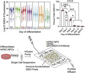
Teratoma formation remains a safety concern in therapeutic cells derived from human-induced pluripotent stem cells (hiPSCs). Residual Teratoma forming cells are present in small numbers in differentiated hiPSC cultures and yet are of significant roadblock to the manufacturing and clinical translation of stem cell therapies. Rare cells are often difficult to remove with standard flow cytometry or magnetic bead sorting techniques. Here, we first characterized time-dependent expression of a teratoma marker, stage-specific embryonic antigen (SSEA)-5, which binds the H type-1 glycan during neural differentiation of hiPSCs. The fraction of cells SSEA-5+ remained high at 97% on day 3, dropped to 70% on day 4, 40% by day 6, and down to 1% on day 12 of differentiation, indicating successful differentiation. We engineered a microfluidic geometrically enhanced differential immunocapture (GEDI) technology to remove SSEA-5+ rare cells from hiPSC-derived neural progenitor cells (hiPSC-NPCs). The GEDI chip removed more than 95% of teratoma-forming cells and presents a facile tool to potentially functionalize with multiple antibodies and robustly enhance hiPSC-derived cell population safety prior to therapeutic transplantation. The approach is potentially amenable to generate a wide variety of high-quality therapeutic cells and can be integrated within the pipeline of cell manufacturing to improve patient safety and reduce the cost of manufacturing through early removal of undesirable cell types.

 求助内容:
求助内容: 应助结果提醒方式:


