Fumiya Nakayama, Makoto Miyoshi, Ai Kimoto, Akari Kawano, Kumiko Miyashita, Shingo Kamoshida, Kazuya Shimizu, Yuichi Hori
{"title":"胰腺癌细胞衍生的外泌体部分通过转化生长因子β1诱导人胰腺癌细胞的上皮-间质转化。","authors":"Fumiya Nakayama, Makoto Miyoshi, Ai Kimoto, Akari Kawano, Kumiko Miyashita, Shingo Kamoshida, Kazuya Shimizu, Yuichi Hori","doi":"10.1007/s00795-022-00321-0","DOIUrl":null,"url":null,"abstract":"<p><p>Distant metastasis is a dismal prognostic factor of pancreatic cancer. Metastasis is established in several steps, but the mechanism underlying the very early stages remains unclear. Epithelial-mesenchymal transition (EMT) is involved in these stages. Although signaling molecules have been reported to induce EMT, the mechanism underlying their origin is unclear. In this study, we hypothesized that pancreatic cancer cell-derived exosomes induce EMT in cancer cells themselves, a notion we entertained because we found EMT in in vitro three-dimensional colonies of cancer cells, with vimentin-positive cells observed in some of the budding pancreatic cancer cells and in single cells outside the colony as well. First, we clarified that pancreatic cancer cell-derived exosomes induce EMT in cancer cells themselves. Next, we examined the involvement of transforming growth factor-β1 (TGF-β1), and TGF-β1 knock-down in pancreatic cancer cells with TGF-β1 siRNA significantly suppressed TGF-β1 gene expression in cancer cells, and exosomal TGF-β1 was significantly reduced in the secretory exosomes. Exosomes from TGF-β1 knock-down cells suppressed EMT induction in cancer cells themselves and TGF-β1 protein expression in target cells. Taken together, these findings suggest that TGF-β1 is involved in EMT induction via exosomes, results that may support the production of effective metastasis inhibitors.</p>","PeriodicalId":29747,"journal":{"name":"Health and History","volume":"16 1","pages":"227-235"},"PeriodicalIF":0.1000,"publicationDate":"2022-09-01","publicationTypes":"Journal Article","fieldsOfStudy":null,"isOpenAccess":false,"openAccessPdf":"https://www.ncbi.nlm.nih.gov/pmc/articles/PMC9043512/pdf/","citationCount":"5","resultStr":"{\"title\":\"Pancreatic cancer cell-derived exosomes induce epithelial-mesenchymal transition in human pancreatic cancer cells themselves partially via transforming growth factor β1.\",\"authors\":\"Fumiya Nakayama, Makoto Miyoshi, Ai Kimoto, Akari Kawano, Kumiko Miyashita, Shingo Kamoshida, Kazuya Shimizu, Yuichi Hori\",\"doi\":\"10.1007/s00795-022-00321-0\",\"DOIUrl\":null,\"url\":null,\"abstract\":\"<p><p>Distant metastasis is a dismal prognostic factor of pancreatic cancer. Metastasis is established in several steps, but the mechanism underlying the very early stages remains unclear. Epithelial-mesenchymal transition (EMT) is involved in these stages. Although signaling molecules have been reported to induce EMT, the mechanism underlying their origin is unclear. In this study, we hypothesized that pancreatic cancer cell-derived exosomes induce EMT in cancer cells themselves, a notion we entertained because we found EMT in in vitro three-dimensional colonies of cancer cells, with vimentin-positive cells observed in some of the budding pancreatic cancer cells and in single cells outside the colony as well. First, we clarified that pancreatic cancer cell-derived exosomes induce EMT in cancer cells themselves. Next, we examined the involvement of transforming growth factor-β1 (TGF-β1), and TGF-β1 knock-down in pancreatic cancer cells with TGF-β1 siRNA significantly suppressed TGF-β1 gene expression in cancer cells, and exosomal TGF-β1 was significantly reduced in the secretory exosomes. Exosomes from TGF-β1 knock-down cells suppressed EMT induction in cancer cells themselves and TGF-β1 protein expression in target cells. Taken together, these findings suggest that TGF-β1 is involved in EMT induction via exosomes, results that may support the production of effective metastasis inhibitors.</p>\",\"PeriodicalId\":29747,\"journal\":{\"name\":\"Health and History\",\"volume\":\"16 1\",\"pages\":\"227-235\"},\"PeriodicalIF\":0.1000,\"publicationDate\":\"2022-09-01\",\"publicationTypes\":\"Journal Article\",\"fieldsOfStudy\":null,\"isOpenAccess\":false,\"openAccessPdf\":\"https://www.ncbi.nlm.nih.gov/pmc/articles/PMC9043512/pdf/\",\"citationCount\":\"5\",\"resultStr\":null,\"platform\":\"Semanticscholar\",\"paperid\":null,\"PeriodicalName\":\"Health and History\",\"FirstCategoryId\":\"3\",\"ListUrlMain\":\"https://doi.org/10.1007/s00795-022-00321-0\",\"RegionNum\":0,\"RegionCategory\":null,\"ArticlePicture\":[],\"TitleCN\":null,\"AbstractTextCN\":null,\"PMCID\":null,\"EPubDate\":\"2022/4/27 0:00:00\",\"PubModel\":\"Epub\",\"JCR\":\"Q3\",\"JCRName\":\"HISTORY & PHILOSOPHY OF SCIENCE\",\"Score\":null,\"Total\":0}","platform":"Semanticscholar","paperid":null,"PeriodicalName":"Health and History","FirstCategoryId":"3","ListUrlMain":"https://doi.org/10.1007/s00795-022-00321-0","RegionNum":0,"RegionCategory":null,"ArticlePicture":[],"TitleCN":null,"AbstractTextCN":null,"PMCID":null,"EPubDate":"2022/4/27 0:00:00","PubModel":"Epub","JCR":"Q3","JCRName":"HISTORY & PHILOSOPHY OF SCIENCE","Score":null,"Total":0}
Pancreatic cancer cell-derived exosomes induce epithelial-mesenchymal transition in human pancreatic cancer cells themselves partially via transforming growth factor β1.
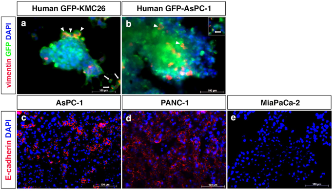
Distant metastasis is a dismal prognostic factor of pancreatic cancer. Metastasis is established in several steps, but the mechanism underlying the very early stages remains unclear. Epithelial-mesenchymal transition (EMT) is involved in these stages. Although signaling molecules have been reported to induce EMT, the mechanism underlying their origin is unclear. In this study, we hypothesized that pancreatic cancer cell-derived exosomes induce EMT in cancer cells themselves, a notion we entertained because we found EMT in in vitro three-dimensional colonies of cancer cells, with vimentin-positive cells observed in some of the budding pancreatic cancer cells and in single cells outside the colony as well. First, we clarified that pancreatic cancer cell-derived exosomes induce EMT in cancer cells themselves. Next, we examined the involvement of transforming growth factor-β1 (TGF-β1), and TGF-β1 knock-down in pancreatic cancer cells with TGF-β1 siRNA significantly suppressed TGF-β1 gene expression in cancer cells, and exosomal TGF-β1 was significantly reduced in the secretory exosomes. Exosomes from TGF-β1 knock-down cells suppressed EMT induction in cancer cells themselves and TGF-β1 protein expression in target cells. Taken together, these findings suggest that TGF-β1 is involved in EMT induction via exosomes, results that may support the production of effective metastasis inhibitors.

 求助内容:
求助内容: 应助结果提醒方式:


