Ali Esmaeil, Ali Ali, Salman Almutairi, Khaled Alkandari, Raed Behbehani, Alaa Alali
{"title":"先天性视盘凹陷和视盘凹陷黄斑病变:综述。","authors":"Ali Esmaeil, Ali Ali, Salman Almutairi, Khaled Alkandari, Raed Behbehani, Alaa Alali","doi":"10.3389/fopht.2023.1222979","DOIUrl":null,"url":null,"abstract":"<p><p>Optic disc pits are a rare but significant anomaly of the optic nerve head that can lead to visual impairment and associated complications. These pits are characterized by a small, oval-shaped depression in the disc, which can cause fluid accumulation and subsequent damage to the adjacent retina. Although the etiology and pathogenesis of optic disc pits are not fully understood, several theories have been proposed, including abnormal embryonic development and degenerative changes. Diagnosis is typically made through a comprehensive eye examination, including a dilated fundus exam and optical coherence tomography. Management options vary depending on the severity of the condition and associated complications, ranging from observation to surgical intervention.</p>","PeriodicalId":13118,"journal":{"name":"Human and Ecological Risk Assessment","volume":"5 1","pages":"1222979"},"PeriodicalIF":2.7000,"publicationDate":"2023-08-16","publicationTypes":"Journal Article","fieldsOfStudy":null,"isOpenAccess":false,"openAccessPdf":"https://www.ncbi.nlm.nih.gov/pmc/articles/PMC11182116/pdf/","citationCount":"0","resultStr":"{\"title\":\"Congenital optic disc pits and optic disc pit maculopathy: a review.\",\"authors\":\"Ali Esmaeil, Ali Ali, Salman Almutairi, Khaled Alkandari, Raed Behbehani, Alaa Alali\",\"doi\":\"10.3389/fopht.2023.1222979\",\"DOIUrl\":null,\"url\":null,\"abstract\":\"<p><p>Optic disc pits are a rare but significant anomaly of the optic nerve head that can lead to visual impairment and associated complications. These pits are characterized by a small, oval-shaped depression in the disc, which can cause fluid accumulation and subsequent damage to the adjacent retina. Although the etiology and pathogenesis of optic disc pits are not fully understood, several theories have been proposed, including abnormal embryonic development and degenerative changes. Diagnosis is typically made through a comprehensive eye examination, including a dilated fundus exam and optical coherence tomography. Management options vary depending on the severity of the condition and associated complications, ranging from observation to surgical intervention.</p>\",\"PeriodicalId\":13118,\"journal\":{\"name\":\"Human and Ecological Risk Assessment\",\"volume\":\"5 1\",\"pages\":\"1222979\"},\"PeriodicalIF\":2.7000,\"publicationDate\":\"2023-08-16\",\"publicationTypes\":\"Journal Article\",\"fieldsOfStudy\":null,\"isOpenAccess\":false,\"openAccessPdf\":\"https://www.ncbi.nlm.nih.gov/pmc/articles/PMC11182116/pdf/\",\"citationCount\":\"0\",\"resultStr\":null,\"platform\":\"Semanticscholar\",\"paperid\":null,\"PeriodicalName\":\"Human and Ecological Risk Assessment\",\"FirstCategoryId\":\"1085\",\"ListUrlMain\":\"https://doi.org/10.3389/fopht.2023.1222979\",\"RegionNum\":3,\"RegionCategory\":\"环境科学与生态学\",\"ArticlePicture\":[],\"TitleCN\":null,\"AbstractTextCN\":null,\"PMCID\":null,\"EPubDate\":\"2023/1/1 0:00:00\",\"PubModel\":\"eCollection\",\"JCR\":\"Q2\",\"JCRName\":\"ENVIRONMENTAL SCIENCES\",\"Score\":null,\"Total\":0}","platform":"Semanticscholar","paperid":null,"PeriodicalName":"Human and Ecological Risk Assessment","FirstCategoryId":"1085","ListUrlMain":"https://doi.org/10.3389/fopht.2023.1222979","RegionNum":3,"RegionCategory":"环境科学与生态学","ArticlePicture":[],"TitleCN":null,"AbstractTextCN":null,"PMCID":null,"EPubDate":"2023/1/1 0:00:00","PubModel":"eCollection","JCR":"Q2","JCRName":"ENVIRONMENTAL SCIENCES","Score":null,"Total":0}
Congenital optic disc pits and optic disc pit maculopathy: a review.
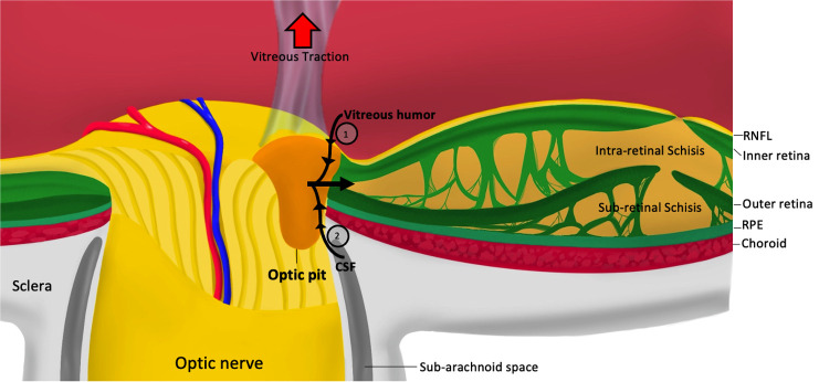
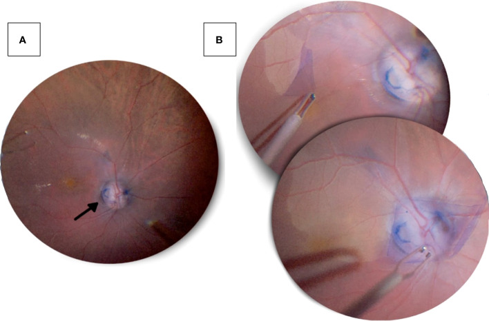
Optic disc pits are a rare but significant anomaly of the optic nerve head that can lead to visual impairment and associated complications. These pits are characterized by a small, oval-shaped depression in the disc, which can cause fluid accumulation and subsequent damage to the adjacent retina. Although the etiology and pathogenesis of optic disc pits are not fully understood, several theories have been proposed, including abnormal embryonic development and degenerative changes. Diagnosis is typically made through a comprehensive eye examination, including a dilated fundus exam and optical coherence tomography. Management options vary depending on the severity of the condition and associated complications, ranging from observation to surgical intervention.
期刊介绍:
Human and Ecological Risk Assessment provides a resource for professionals researching and assessing environmental hazards to both humans and ecological systems. The editors expect papers published to be original, of sound science, purposeful for risk analysis (assessment, communication, management) and related areas, well written (in English), and a contribution to the scientific literature.
The journal''s emphasis is on publication of papers that contribute to improvements in human and ecological health. The journal is an international, fully peer-reviewed publication that publishes eight issues annually. The journal''s scope includes scientific and technical information and critical analysis in the following areas:
-Quantitative Risk Assessment-
Comparative Risk Assessment-
Integrated Human & Ecological Risk Assessment-
Risk Assessment Applications to Human & Ecosystems Health-
Exposure Assessment-
Environmental Fate Assessment-
Multi-Media Assessment-
Hazard Assessment-
Environmental Epidemiology-
Statistical Models and Methods-
Methods Development/Improvement-
Toxicokinetics Modeling-
Animal to Human Extrapolation-
Risk Perception/Communication-
Risk Management-
Regulatory Issues
 求助内容:
求助内容: 应助结果提醒方式:


