Vinny Sara Varghese, John V George, Sylvia Mathew, Shruthi Nagaraja, H N Indiresha, K S Madhu
{"title":"锥形束计算机断层扫描评估两种进洞设计和器械对下颌前牙颈周牙本质厚度的影响。","authors":"Vinny Sara Varghese, John V George, Sylvia Mathew, Shruthi Nagaraja, H N Indiresha, K S Madhu","doi":"10.4103/0972-0707.190018","DOIUrl":null,"url":null,"abstract":"<p><strong>Background and objectives: </strong>The aim of the study was to determine the effect of two access cavity designs on the peri-cervical dentin thickness before and after instrumentation using cone beam computed tomography (CBCT).</p><p><strong>Materials and methods: </strong>Sixty mandibular anterior teeth were divided into two groups of thirty teeth each: Group I: conventional access cavity preparation, where access was prepared just above the cingulum and Group II: incisal access cavity preparation, where access was prepared in proximity to the incisal edge. CBCT scans were taken preoperatively, following access cavity preparation and post instrumentation. 200 μm thick slices were obtained 4mm apical and coronal to the cemento-enamel junction. The peri-cervical dentin thickness was calculated on the facial, lingual, mesial, and distal for all the three obtained scans.</p><p><strong>Results: </strong>The analysis showed that access cavity preparation and instrumentation resulted in a significant loss of tooth structure in Group I on all surfaces, but in Group II, there was a significant loss of tooth structure only in the mesial, lingual, and distal surfaces (P < 0.05).</p><p><strong>Conclusion: </strong>Incisal access cavity preparation resulted in lesser loss of dentin in the peri-cervical region.</p>","PeriodicalId":80359,"journal":{"name":"Advances in cryogenic engineering","volume":"613 1","pages":"450-4"},"PeriodicalIF":0.0000,"publicationDate":"2016-09-01","publicationTypes":"Journal Article","fieldsOfStudy":null,"isOpenAccess":false,"openAccessPdf":"https://www.ncbi.nlm.nih.gov/pmc/articles/PMC5026106/pdf/","citationCount":"0","resultStr":"{\"title\":\"Cone beam computed tomographic evaluation of two access cavity designs and instrumentation on the thickness of peri-cervical dentin in mandibular anterior teeth.\",\"authors\":\"Vinny Sara Varghese, John V George, Sylvia Mathew, Shruthi Nagaraja, H N Indiresha, K S Madhu\",\"doi\":\"10.4103/0972-0707.190018\",\"DOIUrl\":null,\"url\":null,\"abstract\":\"<p><strong>Background and objectives: </strong>The aim of the study was to determine the effect of two access cavity designs on the peri-cervical dentin thickness before and after instrumentation using cone beam computed tomography (CBCT).</p><p><strong>Materials and methods: </strong>Sixty mandibular anterior teeth were divided into two groups of thirty teeth each: Group I: conventional access cavity preparation, where access was prepared just above the cingulum and Group II: incisal access cavity preparation, where access was prepared in proximity to the incisal edge. CBCT scans were taken preoperatively, following access cavity preparation and post instrumentation. 200 μm thick slices were obtained 4mm apical and coronal to the cemento-enamel junction. The peri-cervical dentin thickness was calculated on the facial, lingual, mesial, and distal for all the three obtained scans.</p><p><strong>Results: </strong>The analysis showed that access cavity preparation and instrumentation resulted in a significant loss of tooth structure in Group I on all surfaces, but in Group II, there was a significant loss of tooth structure only in the mesial, lingual, and distal surfaces (P < 0.05).</p><p><strong>Conclusion: </strong>Incisal access cavity preparation resulted in lesser loss of dentin in the peri-cervical region.</p>\",\"PeriodicalId\":80359,\"journal\":{\"name\":\"Advances in cryogenic engineering\",\"volume\":\"613 1\",\"pages\":\"450-4\"},\"PeriodicalIF\":0.0000,\"publicationDate\":\"2016-09-01\",\"publicationTypes\":\"Journal Article\",\"fieldsOfStudy\":null,\"isOpenAccess\":false,\"openAccessPdf\":\"https://www.ncbi.nlm.nih.gov/pmc/articles/PMC5026106/pdf/\",\"citationCount\":\"0\",\"resultStr\":null,\"platform\":\"Semanticscholar\",\"paperid\":null,\"PeriodicalName\":\"Advances in cryogenic engineering\",\"FirstCategoryId\":\"1085\",\"ListUrlMain\":\"https://doi.org/10.4103/0972-0707.190018\",\"RegionNum\":0,\"RegionCategory\":null,\"ArticlePicture\":[],\"TitleCN\":null,\"AbstractTextCN\":null,\"PMCID\":null,\"EPubDate\":\"\",\"PubModel\":\"\",\"JCR\":\"\",\"JCRName\":\"\",\"Score\":null,\"Total\":0}","platform":"Semanticscholar","paperid":null,"PeriodicalName":"Advances in cryogenic engineering","FirstCategoryId":"1085","ListUrlMain":"https://doi.org/10.4103/0972-0707.190018","RegionNum":0,"RegionCategory":null,"ArticlePicture":[],"TitleCN":null,"AbstractTextCN":null,"PMCID":null,"EPubDate":"","PubModel":"","JCR":"","JCRName":"","Score":null,"Total":0}
Cone beam computed tomographic evaluation of two access cavity designs and instrumentation on the thickness of peri-cervical dentin in mandibular anterior teeth.
Background and objectives: The aim of the study was to determine the effect of two access cavity designs on the peri-cervical dentin thickness before and after instrumentation using cone beam computed tomography (CBCT).
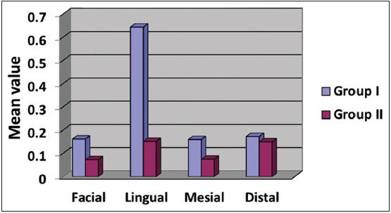
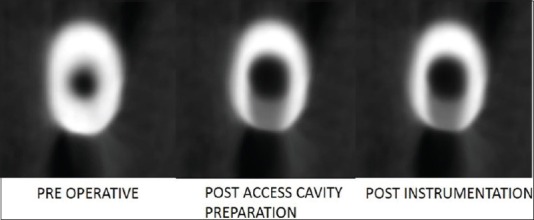
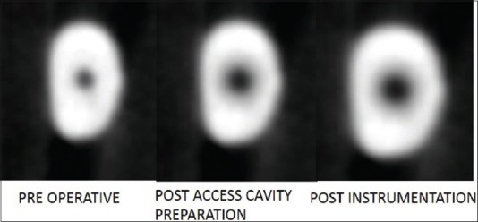
Materials and methods: Sixty mandibular anterior teeth were divided into two groups of thirty teeth each: Group I: conventional access cavity preparation, where access was prepared just above the cingulum and Group II: incisal access cavity preparation, where access was prepared in proximity to the incisal edge. CBCT scans were taken preoperatively, following access cavity preparation and post instrumentation. 200 μm thick slices were obtained 4mm apical and coronal to the cemento-enamel junction. The peri-cervical dentin thickness was calculated on the facial, lingual, mesial, and distal for all the three obtained scans.
Results: The analysis showed that access cavity preparation and instrumentation resulted in a significant loss of tooth structure in Group I on all surfaces, but in Group II, there was a significant loss of tooth structure only in the mesial, lingual, and distal surfaces (P < 0.05).
Conclusion: Incisal access cavity preparation resulted in lesser loss of dentin in the peri-cervical region.

 求助内容:
求助内容: 应助结果提醒方式:


