Alastair J Moss, Mhairi K Doris, Jack P M Andrews, Rong Bing, Marwa Daghem, Edwin J R van Beek, Laura Forsyth, Anoop S V Shah, Michelle C Williams, Stephanie Sellers, Jonathon Leipsic, Marc R Dweck, Richard A Parker, David E Newby, Philip D Adamson
{"title":"18f -氟化物在冠状动脉斑块分子成像中的应用","authors":"Alastair J Moss, Mhairi K Doris, Jack P M Andrews, Rong Bing, Marwa Daghem, Edwin J R van Beek, Laura Forsyth, Anoop S V Shah, Michelle C Williams, Stephanie Sellers, Jonathon Leipsic, Marc R Dweck, Richard A Parker, David E Newby, Philip D Adamson","doi":"10.1161/CIRCIMAGING.118.008574","DOIUrl":null,"url":null,"abstract":"<p><strong>Background: </strong>Coronary <sup>18</sup>F-fluoride positron emission tomography identifies ruptured and high-risk atherosclerotic plaque. The optimal method to identify, to quantify, and to categorize increased coronary <sup>18</sup>F-fluoride uptake and determine its reproducibility has yet to be established. This study aimed to optimize the identification, quantification, categorization, and scan-rescan reproducibility of increased <sup>18</sup>F-fluoride activity in coronary atherosclerotic plaque.</p><p><strong>Methods: </strong>In a prospective observational study, patients with multi-vessel coronary artery disease underwent serial <sup>18</sup>F-fluoride positron emission tomography. Coronary <sup>18</sup>F-fluoride activity was visually assessed, quantified, and categorized with reference to maximal tissue to background ratios. Levels of agreement for both visual and quantitative methods were determined between scans and observers.</p><p><strong>Results: </strong>Thirty patients (90% male, 20 patients with stable coronary artery disease, and 10 with recent type 1 myocardial infarction) underwent paired serial positron emission tomography-coronary computed tomography angiography imaging within an interval of 12±5 days. A mean of 3.7±1.8 <sup>18</sup>F-fluoride positive plaques per patient was identified after recent acute coronary syndrome, compared with 2.4±2.3 positive plaques per patient in stable coronary artery disease. The bias in agreement in maximum tissue to background ratio measurements in visually positive plaques was low between observers (mean difference, -0.01; 95% limits of agreement, -0.32 to 0.30) or between scans (mean difference, 0.06; 95% limits of agreement, -0.49 to 0.61). Good agreement in the categorization of focal <sup>18</sup>F-fluoride uptake was achieved using visual assessment alone (κ=0.66) and further improved at higher maximum tissue to background ratio values.</p><p><strong>Conclusions: </strong>Coronary <sup>18</sup>F-fluoride activity is a precise and reproducible metric in the coronary vasculature. The analytical performance of <sup>18</sup>F-fluoride is sufficient to assess the prognostic utility of this radiotracer as a noninvasive imaging biomarker of plaque vulnerability.</p><p><strong>Clinical trial registration: </strong>URL: http://www.clinicaltrials.gov. Unique identifiers: NCT02110303 and NCT02278211.</p>","PeriodicalId":19614,"journal":{"name":"Origins of Life and Evolution of Biospheres","volume":"25 1","pages":"e008574"},"PeriodicalIF":1.2000,"publicationDate":"2019-08-01","publicationTypes":"Journal Article","fieldsOfStudy":null,"isOpenAccess":false,"openAccessPdf":"https://www.ncbi.nlm.nih.gov/pmc/articles/PMC7668410/pdf/","citationCount":"0","resultStr":"{\"title\":\"Molecular Coronary Plaque Imaging Using <sup>18</sup>F-Fluoride.\",\"authors\":\"Alastair J Moss, Mhairi K Doris, Jack P M Andrews, Rong Bing, Marwa Daghem, Edwin J R van Beek, Laura Forsyth, Anoop S V Shah, Michelle C Williams, Stephanie Sellers, Jonathon Leipsic, Marc R Dweck, Richard A Parker, David E Newby, Philip D Adamson\",\"doi\":\"10.1161/CIRCIMAGING.118.008574\",\"DOIUrl\":null,\"url\":null,\"abstract\":\"<p><strong>Background: </strong>Coronary <sup>18</sup>F-fluoride positron emission tomography identifies ruptured and high-risk atherosclerotic plaque. The optimal method to identify, to quantify, and to categorize increased coronary <sup>18</sup>F-fluoride uptake and determine its reproducibility has yet to be established. This study aimed to optimize the identification, quantification, categorization, and scan-rescan reproducibility of increased <sup>18</sup>F-fluoride activity in coronary atherosclerotic plaque.</p><p><strong>Methods: </strong>In a prospective observational study, patients with multi-vessel coronary artery disease underwent serial <sup>18</sup>F-fluoride positron emission tomography. Coronary <sup>18</sup>F-fluoride activity was visually assessed, quantified, and categorized with reference to maximal tissue to background ratios. Levels of agreement for both visual and quantitative methods were determined between scans and observers.</p><p><strong>Results: </strong>Thirty patients (90% male, 20 patients with stable coronary artery disease, and 10 with recent type 1 myocardial infarction) underwent paired serial positron emission tomography-coronary computed tomography angiography imaging within an interval of 12±5 days. A mean of 3.7±1.8 <sup>18</sup>F-fluoride positive plaques per patient was identified after recent acute coronary syndrome, compared with 2.4±2.3 positive plaques per patient in stable coronary artery disease. The bias in agreement in maximum tissue to background ratio measurements in visually positive plaques was low between observers (mean difference, -0.01; 95% limits of agreement, -0.32 to 0.30) or between scans (mean difference, 0.06; 95% limits of agreement, -0.49 to 0.61). Good agreement in the categorization of focal <sup>18</sup>F-fluoride uptake was achieved using visual assessment alone (κ=0.66) and further improved at higher maximum tissue to background ratio values.</p><p><strong>Conclusions: </strong>Coronary <sup>18</sup>F-fluoride activity is a precise and reproducible metric in the coronary vasculature. The analytical performance of <sup>18</sup>F-fluoride is sufficient to assess the prognostic utility of this radiotracer as a noninvasive imaging biomarker of plaque vulnerability.</p><p><strong>Clinical trial registration: </strong>URL: http://www.clinicaltrials.gov. Unique identifiers: NCT02110303 and NCT02278211.</p>\",\"PeriodicalId\":19614,\"journal\":{\"name\":\"Origins of Life and Evolution of Biospheres\",\"volume\":\"25 1\",\"pages\":\"e008574\"},\"PeriodicalIF\":1.2000,\"publicationDate\":\"2019-08-01\",\"publicationTypes\":\"Journal Article\",\"fieldsOfStudy\":null,\"isOpenAccess\":false,\"openAccessPdf\":\"https://www.ncbi.nlm.nih.gov/pmc/articles/PMC7668410/pdf/\",\"citationCount\":\"0\",\"resultStr\":null,\"platform\":\"Semanticscholar\",\"paperid\":null,\"PeriodicalName\":\"Origins of Life and Evolution of Biospheres\",\"FirstCategoryId\":\"101\",\"ListUrlMain\":\"https://doi.org/10.1161/CIRCIMAGING.118.008574\",\"RegionNum\":4,\"RegionCategory\":\"物理与天体物理\",\"ArticlePicture\":[],\"TitleCN\":null,\"AbstractTextCN\":null,\"PMCID\":null,\"EPubDate\":\"2019/8/6 0:00:00\",\"PubModel\":\"Epub\",\"JCR\":\"Q2\",\"JCRName\":\"BIOLOGY\",\"Score\":null,\"Total\":0}","platform":"Semanticscholar","paperid":null,"PeriodicalName":"Origins of Life and Evolution of Biospheres","FirstCategoryId":"101","ListUrlMain":"https://doi.org/10.1161/CIRCIMAGING.118.008574","RegionNum":4,"RegionCategory":"物理与天体物理","ArticlePicture":[],"TitleCN":null,"AbstractTextCN":null,"PMCID":null,"EPubDate":"2019/8/6 0:00:00","PubModel":"Epub","JCR":"Q2","JCRName":"BIOLOGY","Score":null,"Total":0}
Molecular Coronary Plaque Imaging Using 18F-Fluoride.
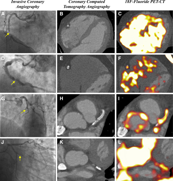
Background: Coronary 18F-fluoride positron emission tomography identifies ruptured and high-risk atherosclerotic plaque. The optimal method to identify, to quantify, and to categorize increased coronary 18F-fluoride uptake and determine its reproducibility has yet to be established. This study aimed to optimize the identification, quantification, categorization, and scan-rescan reproducibility of increased 18F-fluoride activity in coronary atherosclerotic plaque.
Methods: In a prospective observational study, patients with multi-vessel coronary artery disease underwent serial 18F-fluoride positron emission tomography. Coronary 18F-fluoride activity was visually assessed, quantified, and categorized with reference to maximal tissue to background ratios. Levels of agreement for both visual and quantitative methods were determined between scans and observers.
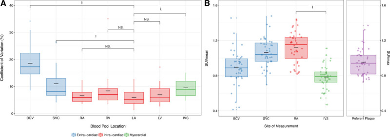
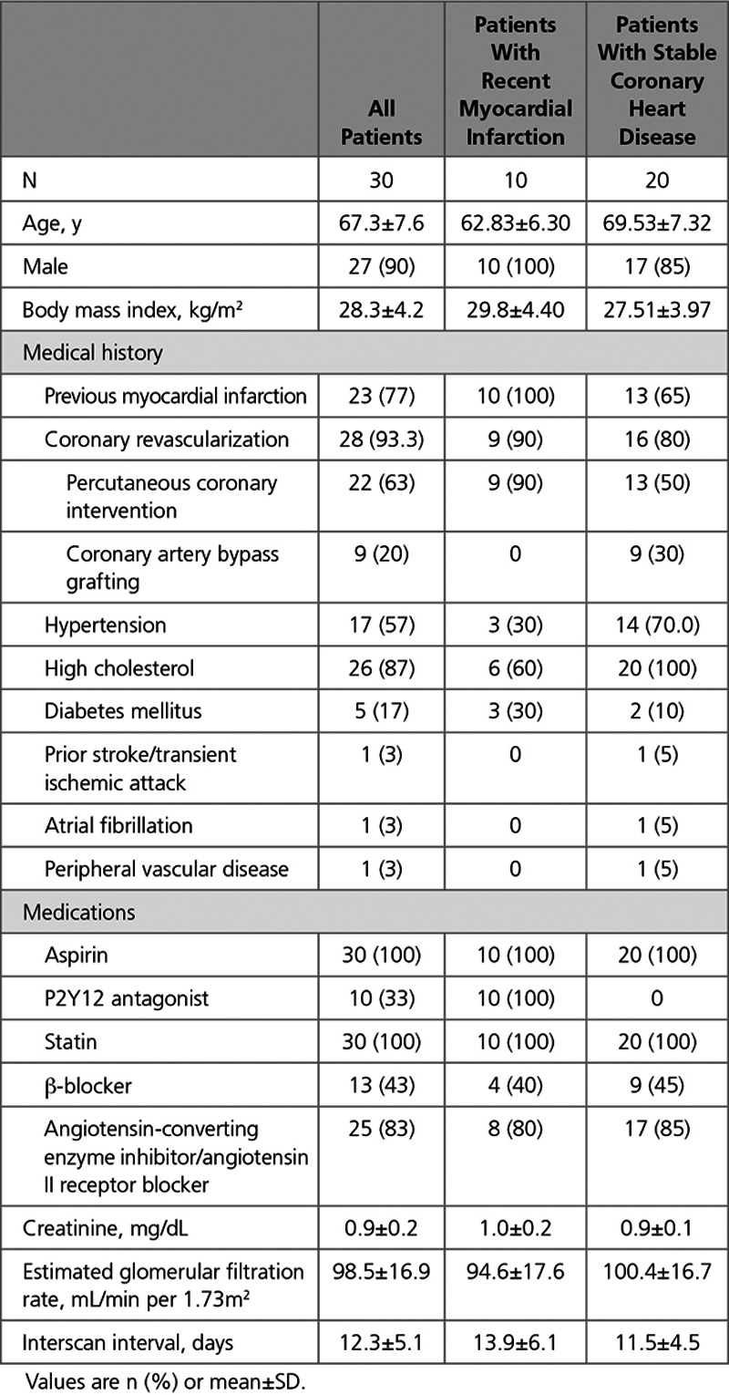
Results: Thirty patients (90% male, 20 patients with stable coronary artery disease, and 10 with recent type 1 myocardial infarction) underwent paired serial positron emission tomography-coronary computed tomography angiography imaging within an interval of 12±5 days. A mean of 3.7±1.8 18F-fluoride positive plaques per patient was identified after recent acute coronary syndrome, compared with 2.4±2.3 positive plaques per patient in stable coronary artery disease. The bias in agreement in maximum tissue to background ratio measurements in visually positive plaques was low between observers (mean difference, -0.01; 95% limits of agreement, -0.32 to 0.30) or between scans (mean difference, 0.06; 95% limits of agreement, -0.49 to 0.61). Good agreement in the categorization of focal 18F-fluoride uptake was achieved using visual assessment alone (κ=0.66) and further improved at higher maximum tissue to background ratio values.
Conclusions: Coronary 18F-fluoride activity is a precise and reproducible metric in the coronary vasculature. The analytical performance of 18F-fluoride is sufficient to assess the prognostic utility of this radiotracer as a noninvasive imaging biomarker of plaque vulnerability.
Clinical trial registration: URL: http://www.clinicaltrials.gov. Unique identifiers: NCT02110303 and NCT02278211.
期刊介绍:
The subject of the origin and early evolution of life is an inseparable part of the general discipline of Astrobiology. The journal Origins of Life and Evolution of Biospheres places special importance on the interconnection as well as the interdisciplinary nature of these fields, as is reflected in its subject coverage. While any scientific study which contributes to our understanding of the origins, evolution and distribution of life in the Universe is suitable for inclusion in the journal, some examples of important areas of interest are: prebiotic chemistry and the nature of Earth''s early environment, self-replicating and self-organizing systems, the theory of the RNA world and of other possible precursor systems, and the problem of the origin of the genetic code. Early evolution of life - as revealed by such techniques as the elucidation of biochemical pathways, molecular phylogeny, the study of Precambrian sediments and fossils and of major innovations in microbial evolution - forms a second focus. As a larger and more general context for these areas, Astrobiology refers to the origin and evolution of life in a cosmic setting, and includes interstellar chemistry, planetary atmospheres and habitable zones, the organic chemistry of comets, meteorites, asteroids and other small bodies, biological adaptation to extreme environments, life detection and related areas. Experimental papers, theoretical articles and authorative literature reviews are all appropriate forms for submission to the journal. In the coming years, Astrobiology will play an even greater role in defining the journal''s coverage and keeping Origins of Life and Evolution of Biospheres well-placed in this growing interdisciplinary field.

 求助内容:
求助内容: 应助结果提醒方式:


