Shreyas Dudhani, Amit Kumar Sinha, Bindey Kumar, Amit Kumar, Monika Anant
{"title":"伴有尿道重复的尿道窦畸形中的巨大结石:病例报告与文献综述。","authors":"Shreyas Dudhani, Amit Kumar Sinha, Bindey Kumar, Amit Kumar, Monika Anant","doi":"10.4103/ajps.ajps_89_22","DOIUrl":null,"url":null,"abstract":"<p><strong>Abstract: </strong>Primary vaginal calculi are uncommon in children. Urethral duplication in females is seen to occur in association with complex congenital malformations. We report the case of perianal persistent urogenital sinus with a hypertrophied clitoris with phallic urethra, scrotum-like pouch, uterus didelphys with obstructed hemivagina, and giant colpolithiasis in 46XX female. A 16-year-old presented with pain abdomen and cyclic passage of blood clots per rectum. She had a tender lump in left iliac region, a phallus like protrusion and a ruggous sac below it. Vaginal opening was absent. Computed tomography showed two uterine horns with a separate cervix and distended non-communicating hemivaginas with a large calcified oval mass in the left hemivagina. On exploration, calculus was extracted from the left hemivagina. The large calculus found in the left hemivagina appears to be the cause of all presenting symptoms. It obstructed the left hemivagina, filling the left uterine horn with menstrual blood causing its gradual enlargement and secondary infection. The early diagnosis and prompt referral of such an anomaly can only be ensured in institutional deliveries. For a significant proportion of newborns in the developing world, the ability to afford or even be referred to institutes which deal with such cases is a luxurious affair. We hope to bridge bridging the knowledge, attitude and practice gap that exists in our health-care system with this report.</p>","PeriodicalId":72123,"journal":{"name":"African journal of paediatric surgery : AJPS","volume":"1 1","pages":"69-72"},"PeriodicalIF":0.0000,"publicationDate":"2024-01-01","publicationTypes":"Journal Article","fieldsOfStudy":null,"isOpenAccess":false,"openAccessPdf":"https://www.ncbi.nlm.nih.gov/pmc/articles/PMC10903723/pdf/","citationCount":"0","resultStr":"{\"title\":\"Giant Colpolithiasis in Urogenital Sinus Anomaly with Urethral Duplication: A Case Report and Review of Literature.\",\"authors\":\"Shreyas Dudhani, Amit Kumar Sinha, Bindey Kumar, Amit Kumar, Monika Anant\",\"doi\":\"10.4103/ajps.ajps_89_22\",\"DOIUrl\":null,\"url\":null,\"abstract\":\"<p><strong>Abstract: </strong>Primary vaginal calculi are uncommon in children. Urethral duplication in females is seen to occur in association with complex congenital malformations. We report the case of perianal persistent urogenital sinus with a hypertrophied clitoris with phallic urethra, scrotum-like pouch, uterus didelphys with obstructed hemivagina, and giant colpolithiasis in 46XX female. A 16-year-old presented with pain abdomen and cyclic passage of blood clots per rectum. She had a tender lump in left iliac region, a phallus like protrusion and a ruggous sac below it. Vaginal opening was absent. Computed tomography showed two uterine horns with a separate cervix and distended non-communicating hemivaginas with a large calcified oval mass in the left hemivagina. On exploration, calculus was extracted from the left hemivagina. The large calculus found in the left hemivagina appears to be the cause of all presenting symptoms. It obstructed the left hemivagina, filling the left uterine horn with menstrual blood causing its gradual enlargement and secondary infection. The early diagnosis and prompt referral of such an anomaly can only be ensured in institutional deliveries. For a significant proportion of newborns in the developing world, the ability to afford or even be referred to institutes which deal with such cases is a luxurious affair. We hope to bridge bridging the knowledge, attitude and practice gap that exists in our health-care system with this report.</p>\",\"PeriodicalId\":72123,\"journal\":{\"name\":\"African journal of paediatric surgery : AJPS\",\"volume\":\"1 1\",\"pages\":\"69-72\"},\"PeriodicalIF\":0.0000,\"publicationDate\":\"2024-01-01\",\"publicationTypes\":\"Journal Article\",\"fieldsOfStudy\":null,\"isOpenAccess\":false,\"openAccessPdf\":\"https://www.ncbi.nlm.nih.gov/pmc/articles/PMC10903723/pdf/\",\"citationCount\":\"0\",\"resultStr\":null,\"platform\":\"Semanticscholar\",\"paperid\":null,\"PeriodicalName\":\"African journal of paediatric surgery : AJPS\",\"FirstCategoryId\":\"1085\",\"ListUrlMain\":\"https://doi.org/10.4103/ajps.ajps_89_22\",\"RegionNum\":0,\"RegionCategory\":null,\"ArticlePicture\":[],\"TitleCN\":null,\"AbstractTextCN\":null,\"PMCID\":null,\"EPubDate\":\"2023/1/19 0:00:00\",\"PubModel\":\"Epub\",\"JCR\":\"\",\"JCRName\":\"\",\"Score\":null,\"Total\":0}","platform":"Semanticscholar","paperid":null,"PeriodicalName":"African journal of paediatric surgery : AJPS","FirstCategoryId":"1085","ListUrlMain":"https://doi.org/10.4103/ajps.ajps_89_22","RegionNum":0,"RegionCategory":null,"ArticlePicture":[],"TitleCN":null,"AbstractTextCN":null,"PMCID":null,"EPubDate":"2023/1/19 0:00:00","PubModel":"Epub","JCR":"","JCRName":"","Score":null,"Total":0}
Giant Colpolithiasis in Urogenital Sinus Anomaly with Urethral Duplication: A Case Report and Review of Literature.
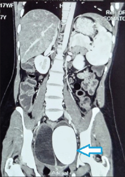
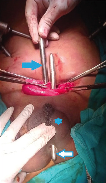
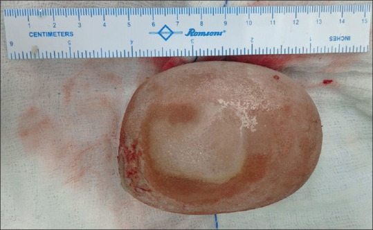
Abstract: Primary vaginal calculi are uncommon in children. Urethral duplication in females is seen to occur in association with complex congenital malformations. We report the case of perianal persistent urogenital sinus with a hypertrophied clitoris with phallic urethra, scrotum-like pouch, uterus didelphys with obstructed hemivagina, and giant colpolithiasis in 46XX female. A 16-year-old presented with pain abdomen and cyclic passage of blood clots per rectum. She had a tender lump in left iliac region, a phallus like protrusion and a ruggous sac below it. Vaginal opening was absent. Computed tomography showed two uterine horns with a separate cervix and distended non-communicating hemivaginas with a large calcified oval mass in the left hemivagina. On exploration, calculus was extracted from the left hemivagina. The large calculus found in the left hemivagina appears to be the cause of all presenting symptoms. It obstructed the left hemivagina, filling the left uterine horn with menstrual blood causing its gradual enlargement and secondary infection. The early diagnosis and prompt referral of such an anomaly can only be ensured in institutional deliveries. For a significant proportion of newborns in the developing world, the ability to afford or even be referred to institutes which deal with such cases is a luxurious affair. We hope to bridge bridging the knowledge, attitude and practice gap that exists in our health-care system with this report.

 求助内容:
求助内容: 应助结果提醒方式:


