Immacolata Tartaglione, Camilla Russo, Andrea Elefante, Martina Caiazza, Maddalena Casale, Rosanna Di Concilio, Angela Ciancio, Elisa De Michele, Giovanni Amendola, Paolo Gritti, Pasquale A. Carafa, Teresa Ferrantino, Antonella Centanni, Noemi Ippolito, Violetta Caserta, Tiziana Oliveto, Ilaria Granato, Gianluca Femina, Fabrizio Esposito, Sara Ponticorvo, Andrea G. Russo, Antonietta Canna, Mario Ermani, Mario Cirillo, Silverio Perrotta, Renzo Manara
{"title":"无证据表明成人无神经症状β-地中海贫血患者脑血管受累增加。多中心多模态磁共振研究","authors":"Immacolata Tartaglione, Camilla Russo, Andrea Elefante, Martina Caiazza, Maddalena Casale, Rosanna Di Concilio, Angela Ciancio, Elisa De Michele, Giovanni Amendola, Paolo Gritti, Pasquale A. Carafa, Teresa Ferrantino, Antonella Centanni, Noemi Ippolito, Violetta Caserta, Tiziana Oliveto, Ilaria Granato, Gianluca Femina, Fabrizio Esposito, Sara Ponticorvo, Andrea G. Russo, Antonietta Canna, Mario Ermani, Mario Cirillo, Silverio Perrotta, Renzo Manara","doi":"10.1111/bjh.15834","DOIUrl":null,"url":null,"abstract":"<p>Multi-factorial causes jeopardize brain integrity in β-thalassaemia. Intracranial parenchymal and vascular changes have been reported among young β-thalassaemia patients but conventional magnetic resonance imaging (MRI) findings are contradictory making early MRI and magnetic resonance angiography (MRA)/venography monitoring a matter of debate.</p><p>This study prospectively investigated 75 neurologically asymptomatic β-thalassaemia patients (mean-age 35·2 ± 10·7 years; 52/75 transfusion-dependent; 41/75 splenectomised) using a 3T magnetic resonance scanner; clinical, laboratory and treatment data were also collected. White matter ischaemic-like abnormalities, intracranial artery stenoses, aneurysms and sinus venous thrombosis were compared between patients and 56 healthy controls (mean-age 33·9 ± 10·8 years). No patient or control showed silent territorial or lacunar strokes, intracranial artery stenoses or signs of sinus thrombosis. White matter lesions were found both in patients (35/75, 46·7%) and controls (28/56, 50·0%), without differences in terms of number (4·0 ± 10·6 vs. 4·6 ± 9·1, <i>P</i> = 0·63), size and Fazekas’ Score. Intracranial aneurysms did not differ between patients and controls for incidence rate (7/75, 9·3% vs. 5/56, 8·9%), size and site. Vascular and parenchymal abnormality rate did not differ according to treatments or clinical phenotype. According to this study, asymptomatic β-thalassaemia patients treated according to current guidelines do not seem to carry an increased risk of brain and intracranial vascular changes, thus weakening recommendations for regular brain MRI monitoring.</p>","PeriodicalId":135,"journal":{"name":"British Journal of Haematology","volume":null,"pages":null},"PeriodicalIF":5.1000,"publicationDate":"2019-03-05","publicationTypes":"Journal Article","fieldsOfStudy":null,"isOpenAccess":false,"openAccessPdf":"https://sci-hub-pdf.com/10.1111/bjh.15834","citationCount":"13","resultStr":"{\"title\":\"No evidence of increased cerebrovascular involvement in adult neurologically-asymptomatic β-Thalassaemia. A multicentre multimodal magnetic resonance study\",\"authors\":\"Immacolata Tartaglione, Camilla Russo, Andrea Elefante, Martina Caiazza, Maddalena Casale, Rosanna Di Concilio, Angela Ciancio, Elisa De Michele, Giovanni Amendola, Paolo Gritti, Pasquale A. Carafa, Teresa Ferrantino, Antonella Centanni, Noemi Ippolito, Violetta Caserta, Tiziana Oliveto, Ilaria Granato, Gianluca Femina, Fabrizio Esposito, Sara Ponticorvo, Andrea G. Russo, Antonietta Canna, Mario Ermani, Mario Cirillo, Silverio Perrotta, Renzo Manara\",\"doi\":\"10.1111/bjh.15834\",\"DOIUrl\":null,\"url\":null,\"abstract\":\"<p>Multi-factorial causes jeopardize brain integrity in β-thalassaemia. Intracranial parenchymal and vascular changes have been reported among young β-thalassaemia patients but conventional magnetic resonance imaging (MRI) findings are contradictory making early MRI and magnetic resonance angiography (MRA)/venography monitoring a matter of debate.</p><p>This study prospectively investigated 75 neurologically asymptomatic β-thalassaemia patients (mean-age 35·2 ± 10·7 years; 52/75 transfusion-dependent; 41/75 splenectomised) using a 3T magnetic resonance scanner; clinical, laboratory and treatment data were also collected. White matter ischaemic-like abnormalities, intracranial artery stenoses, aneurysms and sinus venous thrombosis were compared between patients and 56 healthy controls (mean-age 33·9 ± 10·8 years). No patient or control showed silent territorial or lacunar strokes, intracranial artery stenoses or signs of sinus thrombosis. White matter lesions were found both in patients (35/75, 46·7%) and controls (28/56, 50·0%), without differences in terms of number (4·0 ± 10·6 vs. 4·6 ± 9·1, <i>P</i> = 0·63), size and Fazekas’ Score. Intracranial aneurysms did not differ between patients and controls for incidence rate (7/75, 9·3% vs. 5/56, 8·9%), size and site. Vascular and parenchymal abnormality rate did not differ according to treatments or clinical phenotype. According to this study, asymptomatic β-thalassaemia patients treated according to current guidelines do not seem to carry an increased risk of brain and intracranial vascular changes, thus weakening recommendations for regular brain MRI monitoring.</p>\",\"PeriodicalId\":135,\"journal\":{\"name\":\"British Journal of Haematology\",\"volume\":null,\"pages\":null},\"PeriodicalIF\":5.1000,\"publicationDate\":\"2019-03-05\",\"publicationTypes\":\"Journal Article\",\"fieldsOfStudy\":null,\"isOpenAccess\":false,\"openAccessPdf\":\"https://sci-hub-pdf.com/10.1111/bjh.15834\",\"citationCount\":\"13\",\"resultStr\":null,\"platform\":\"Semanticscholar\",\"paperid\":null,\"PeriodicalName\":\"British Journal of Haematology\",\"FirstCategoryId\":\"3\",\"ListUrlMain\":\"https://onlinelibrary.wiley.com/doi/10.1111/bjh.15834\",\"RegionNum\":2,\"RegionCategory\":\"医学\",\"ArticlePicture\":[],\"TitleCN\":null,\"AbstractTextCN\":null,\"PMCID\":null,\"EPubDate\":\"\",\"PubModel\":\"\",\"JCR\":\"Q1\",\"JCRName\":\"HEMATOLOGY\",\"Score\":null,\"Total\":0}","platform":"Semanticscholar","paperid":null,"PeriodicalName":"British Journal of Haematology","FirstCategoryId":"3","ListUrlMain":"https://onlinelibrary.wiley.com/doi/10.1111/bjh.15834","RegionNum":2,"RegionCategory":"医学","ArticlePicture":[],"TitleCN":null,"AbstractTextCN":null,"PMCID":null,"EPubDate":"","PubModel":"","JCR":"Q1","JCRName":"HEMATOLOGY","Score":null,"Total":0}
引用次数: 13
摘要
β-地中海贫血的多因素原因危及脑完整性。年轻β-地中海贫血患者的颅内实质和血管改变已被报道,但常规磁共振成像(MRI)的发现是矛盾的,这使得早期MRI和磁共振血管造影(MRA)/静脉造影监测成为一个有争议的问题。本研究前瞻性调查了75例神经无症状β-地中海贫血患者(平均年龄35.2±10.7岁;52/75 transfusion-dependent;41/75脾切除术)使用3T磁共振扫描仪;同时收集临床、实验室和治疗资料。比较患者与56名健康对照者(平均年龄33.9±10.8岁)脑白质缺血样异常、颅内动脉狭窄、动脉瘤及窦静脉血栓形成情况。没有患者或对照组表现出无症状的区域性或腔隙性中风,颅内动脉狭窄或窦血栓形成的迹象。患者(35/ 75,46·7%)和对照组(28/ 56,50·0%)均有白质病变,在数量(4·0±10·6 vs. 4·6±9·1,P = 0.63)、大小和Fazekas评分方面无差异。颅内动脉瘤的发生率(7/ 75,9·3% vs. 5/ 56,8·9%)、大小和部位在患者和对照组之间没有差异。血管和实质异常率不因治疗或临床表型而异。根据这项研究,根据现行指南治疗的无症状β-地中海贫血患者似乎没有增加脑和颅内血管改变的风险,因此削弱了定期进行脑MRI监测的建议。
No evidence of increased cerebrovascular involvement in adult neurologically-asymptomatic β-Thalassaemia. A multicentre multimodal magnetic resonance study
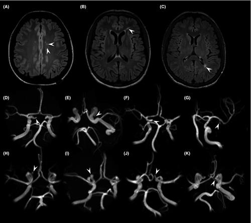
Multi-factorial causes jeopardize brain integrity in β-thalassaemia. Intracranial parenchymal and vascular changes have been reported among young β-thalassaemia patients but conventional magnetic resonance imaging (MRI) findings are contradictory making early MRI and magnetic resonance angiography (MRA)/venography monitoring a matter of debate.
This study prospectively investigated 75 neurologically asymptomatic β-thalassaemia patients (mean-age 35·2 ± 10·7 years; 52/75 transfusion-dependent; 41/75 splenectomised) using a 3T magnetic resonance scanner; clinical, laboratory and treatment data were also collected. White matter ischaemic-like abnormalities, intracranial artery stenoses, aneurysms and sinus venous thrombosis were compared between patients and 56 healthy controls (mean-age 33·9 ± 10·8 years). No patient or control showed silent territorial or lacunar strokes, intracranial artery stenoses or signs of sinus thrombosis. White matter lesions were found both in patients (35/75, 46·7%) and controls (28/56, 50·0%), without differences in terms of number (4·0 ± 10·6 vs. 4·6 ± 9·1, P = 0·63), size and Fazekas’ Score. Intracranial aneurysms did not differ between patients and controls for incidence rate (7/75, 9·3% vs. 5/56, 8·9%), size and site. Vascular and parenchymal abnormality rate did not differ according to treatments or clinical phenotype. According to this study, asymptomatic β-thalassaemia patients treated according to current guidelines do not seem to carry an increased risk of brain and intracranial vascular changes, thus weakening recommendations for regular brain MRI monitoring.
期刊介绍:
The British Journal of Haematology publishes original research papers in clinical, laboratory and experimental haematology. The Journal also features annotations, reviews, short reports, images in haematology and Letters to the Editor.
 求助内容:
求助内容: 应助结果提醒方式:


