{"title":"大面积局部性腹部淋巴水肿:病例报告与文献综述","authors":"Badri Gogia, Irina Chekmareva, Anastasiia Leonova, Rifat Alyautdinov, Grigory Karmazanovsky, Andrey Glotov, Dmitry Kalinin","doi":"10.1055/a-2140-8589","DOIUrl":null,"url":null,"abstract":"<p><p>Massive localized lymphedema (MLL) is a rare disease caused by the obstruction of lymphatic vessels with specific clinical morphological and radiological characteristics. People with morbid obesity are mainly affected by MLL. Lymphedema is easily confused with soft tissue sarcoma and requires differential diagnosis, both the possibility of an MLL and also carcinoma manifestations in the soft tissues. The possible causes of massive lymphedema include trauma, surgery, and hypothyroidism. This report is the first case of MLL treated surgically in the Russian Federation. Detailed computed tomography (CT) characteristics and an electron microscope picture of MLL are discussed. A 50-year-old woman (body mass index of 43 kg/m <sup>2</sup> ) with MLL arising from the anterior abdominal wall was admitted to the hospital for surgical treatment. Its mass was 22.16 kg. A morphological study of the resected mass confirmed the diagnosis of MLL. We review etiology, clinical presentation, diagnosis, and treatment of MLL. We also performed an electron-microscopic study that revealed interstitial Cajal-like cells telocytes not previously described in MLL cases. We did not find similar findings in the literature. It is possible that the conduction of an ultrastructural examination of MLL tissue samples will further contribute to the understanding of MLL pathogenesis.</p>","PeriodicalId":47543,"journal":{"name":"Archives of Plastic Surgery-APS","volume":"1 1","pages":"615-620"},"PeriodicalIF":1.5000,"publicationDate":"2023-11-30","publicationTypes":"Journal Article","fieldsOfStudy":null,"isOpenAccess":false,"openAccessPdf":"https://www.ncbi.nlm.nih.gov/pmc/articles/PMC10736210/pdf/","citationCount":"0","resultStr":"{\"title\":\"Massive Localized Abdominal Lymphedema: A Case Report with Literature Review.\",\"authors\":\"Badri Gogia, Irina Chekmareva, Anastasiia Leonova, Rifat Alyautdinov, Grigory Karmazanovsky, Andrey Glotov, Dmitry Kalinin\",\"doi\":\"10.1055/a-2140-8589\",\"DOIUrl\":null,\"url\":null,\"abstract\":\"<p><p>Massive localized lymphedema (MLL) is a rare disease caused by the obstruction of lymphatic vessels with specific clinical morphological and radiological characteristics. People with morbid obesity are mainly affected by MLL. Lymphedema is easily confused with soft tissue sarcoma and requires differential diagnosis, both the possibility of an MLL and also carcinoma manifestations in the soft tissues. The possible causes of massive lymphedema include trauma, surgery, and hypothyroidism. This report is the first case of MLL treated surgically in the Russian Federation. Detailed computed tomography (CT) characteristics and an electron microscope picture of MLL are discussed. A 50-year-old woman (body mass index of 43 kg/m <sup>2</sup> ) with MLL arising from the anterior abdominal wall was admitted to the hospital for surgical treatment. Its mass was 22.16 kg. A morphological study of the resected mass confirmed the diagnosis of MLL. We review etiology, clinical presentation, diagnosis, and treatment of MLL. We also performed an electron-microscopic study that revealed interstitial Cajal-like cells telocytes not previously described in MLL cases. We did not find similar findings in the literature. It is possible that the conduction of an ultrastructural examination of MLL tissue samples will further contribute to the understanding of MLL pathogenesis.</p>\",\"PeriodicalId\":47543,\"journal\":{\"name\":\"Archives of Plastic Surgery-APS\",\"volume\":\"1 1\",\"pages\":\"615-620\"},\"PeriodicalIF\":1.5000,\"publicationDate\":\"2023-11-30\",\"publicationTypes\":\"Journal Article\",\"fieldsOfStudy\":null,\"isOpenAccess\":false,\"openAccessPdf\":\"https://www.ncbi.nlm.nih.gov/pmc/articles/PMC10736210/pdf/\",\"citationCount\":\"0\",\"resultStr\":null,\"platform\":\"Semanticscholar\",\"paperid\":null,\"PeriodicalName\":\"Archives of Plastic Surgery-APS\",\"FirstCategoryId\":\"1085\",\"ListUrlMain\":\"https://doi.org/10.1055/a-2140-8589\",\"RegionNum\":0,\"RegionCategory\":null,\"ArticlePicture\":[],\"TitleCN\":null,\"AbstractTextCN\":null,\"PMCID\":null,\"EPubDate\":\"2023/11/1 0:00:00\",\"PubModel\":\"eCollection\",\"JCR\":\"Q3\",\"JCRName\":\"SURGERY\",\"Score\":null,\"Total\":0}","platform":"Semanticscholar","paperid":null,"PeriodicalName":"Archives of Plastic Surgery-APS","FirstCategoryId":"1085","ListUrlMain":"https://doi.org/10.1055/a-2140-8589","RegionNum":0,"RegionCategory":null,"ArticlePicture":[],"TitleCN":null,"AbstractTextCN":null,"PMCID":null,"EPubDate":"2023/11/1 0:00:00","PubModel":"eCollection","JCR":"Q3","JCRName":"SURGERY","Score":null,"Total":0}
Massive Localized Abdominal Lymphedema: A Case Report with Literature Review.
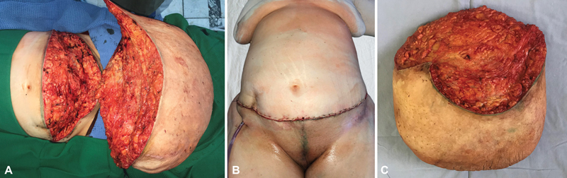
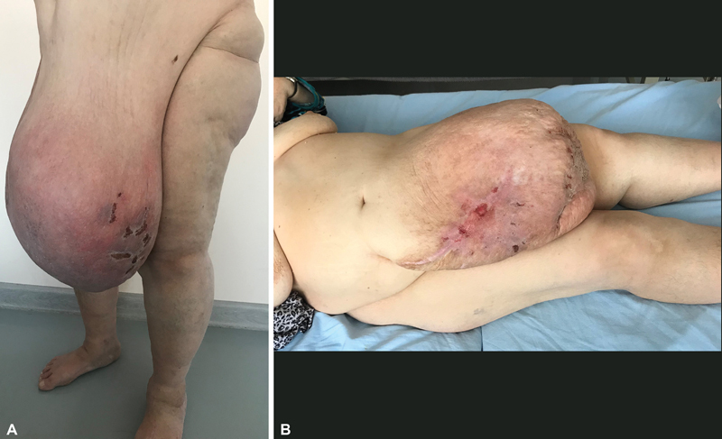
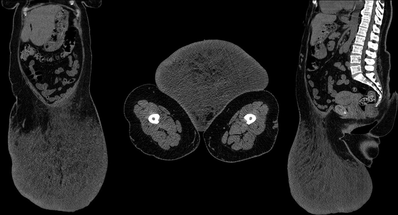
Massive localized lymphedema (MLL) is a rare disease caused by the obstruction of lymphatic vessels with specific clinical morphological and radiological characteristics. People with morbid obesity are mainly affected by MLL. Lymphedema is easily confused with soft tissue sarcoma and requires differential diagnosis, both the possibility of an MLL and also carcinoma manifestations in the soft tissues. The possible causes of massive lymphedema include trauma, surgery, and hypothyroidism. This report is the first case of MLL treated surgically in the Russian Federation. Detailed computed tomography (CT) characteristics and an electron microscope picture of MLL are discussed. A 50-year-old woman (body mass index of 43 kg/m 2 ) with MLL arising from the anterior abdominal wall was admitted to the hospital for surgical treatment. Its mass was 22.16 kg. A morphological study of the resected mass confirmed the diagnosis of MLL. We review etiology, clinical presentation, diagnosis, and treatment of MLL. We also performed an electron-microscopic study that revealed interstitial Cajal-like cells telocytes not previously described in MLL cases. We did not find similar findings in the literature. It is possible that the conduction of an ultrastructural examination of MLL tissue samples will further contribute to the understanding of MLL pathogenesis.

 求助内容:
求助内容: 应助结果提醒方式:


