{"title":"基于弥散张量成像(DTI)的脑白质成像研究综述","authors":"Yaniv Assaf, Ofer Pasternak","doi":"10.1007/s12031-007-0029-0","DOIUrl":null,"url":null,"abstract":"<p>Diffusion tensor imaging (DTI) has become one of the most popular MRI techniques in brain research, as well as in clinical practice. The number of brain studies with DTI is growing steadily and, over the last decade, has produced more than 700 publications. Diffusion tensor imaging enables visualization and characterization of white matter fascicli in two and three dimensions. Since the introduction of this methodology in 1994, it has been used to study the white matter architecture and integrity of the normal and diseased brains (multiple sclerosis, stroke, aging, dementia, schizophrenia, etc.). Although it provided image contrast that was not available with routine MR techniques, unique information on white matter and 3D visualization of neuronal pathways, many questions were raised regarding the origin of the DTI signal. Diffusion tensor imaging is constantly validated, challenged, and developed in terms of acquisition scheme, image processing, analysis, and interpretation. While DTI offers a powerful tool to study and visualize white matter, it suffers from inherent artifacts and limitations. The partial volume effect and the inability of the model to cope with non-Gaussian diffusion are its two main drawbacks. Nevertheless, when combined with functional brain mapping, DTI provides an efficient tool for comprehensive, noninvasive, functional anatomy mapping of the human brain. This review summarizes the development of DTI in the last decade with respect to the specificity and utility of the technique in radiology and anatomy studies.</p>","PeriodicalId":652,"journal":{"name":"Journal of Molecular Neuroscience","volume":"34 1","pages":"51 - 61"},"PeriodicalIF":2.8000,"publicationDate":"2007-09-27","publicationTypes":"Journal Article","fieldsOfStudy":null,"isOpenAccess":false,"openAccessPdf":"https://sci-hub-pdf.com/10.1007/s12031-007-0029-0","citationCount":"1311","resultStr":"{\"title\":\"Diffusion Tensor Imaging (DTI)-based White Matter Mapping in Brain Research: A Review\",\"authors\":\"Yaniv Assaf, Ofer Pasternak\",\"doi\":\"10.1007/s12031-007-0029-0\",\"DOIUrl\":null,\"url\":null,\"abstract\":\"<p>Diffusion tensor imaging (DTI) has become one of the most popular MRI techniques in brain research, as well as in clinical practice. The number of brain studies with DTI is growing steadily and, over the last decade, has produced more than 700 publications. Diffusion tensor imaging enables visualization and characterization of white matter fascicli in two and three dimensions. Since the introduction of this methodology in 1994, it has been used to study the white matter architecture and integrity of the normal and diseased brains (multiple sclerosis, stroke, aging, dementia, schizophrenia, etc.). Although it provided image contrast that was not available with routine MR techniques, unique information on white matter and 3D visualization of neuronal pathways, many questions were raised regarding the origin of the DTI signal. Diffusion tensor imaging is constantly validated, challenged, and developed in terms of acquisition scheme, image processing, analysis, and interpretation. While DTI offers a powerful tool to study and visualize white matter, it suffers from inherent artifacts and limitations. The partial volume effect and the inability of the model to cope with non-Gaussian diffusion are its two main drawbacks. Nevertheless, when combined with functional brain mapping, DTI provides an efficient tool for comprehensive, noninvasive, functional anatomy mapping of the human brain. This review summarizes the development of DTI in the last decade with respect to the specificity and utility of the technique in radiology and anatomy studies.</p>\",\"PeriodicalId\":652,\"journal\":{\"name\":\"Journal of Molecular Neuroscience\",\"volume\":\"34 1\",\"pages\":\"51 - 61\"},\"PeriodicalIF\":2.8000,\"publicationDate\":\"2007-09-27\",\"publicationTypes\":\"Journal Article\",\"fieldsOfStudy\":null,\"isOpenAccess\":false,\"openAccessPdf\":\"https://sci-hub-pdf.com/10.1007/s12031-007-0029-0\",\"citationCount\":\"1311\",\"resultStr\":null,\"platform\":\"Semanticscholar\",\"paperid\":null,\"PeriodicalName\":\"Journal of Molecular Neuroscience\",\"FirstCategoryId\":\"3\",\"ListUrlMain\":\"https://link.springer.com/article/10.1007/s12031-007-0029-0\",\"RegionNum\":4,\"RegionCategory\":\"医学\",\"ArticlePicture\":[],\"TitleCN\":null,\"AbstractTextCN\":null,\"PMCID\":null,\"EPubDate\":\"\",\"PubModel\":\"\",\"JCR\":\"Q3\",\"JCRName\":\"BIOCHEMISTRY & MOLECULAR BIOLOGY\",\"Score\":null,\"Total\":0}","platform":"Semanticscholar","paperid":null,"PeriodicalName":"Journal of Molecular Neuroscience","FirstCategoryId":"3","ListUrlMain":"https://link.springer.com/article/10.1007/s12031-007-0029-0","RegionNum":4,"RegionCategory":"医学","ArticlePicture":[],"TitleCN":null,"AbstractTextCN":null,"PMCID":null,"EPubDate":"","PubModel":"","JCR":"Q3","JCRName":"BIOCHEMISTRY & MOLECULAR BIOLOGY","Score":null,"Total":0}
Diffusion Tensor Imaging (DTI)-based White Matter Mapping in Brain Research: A Review
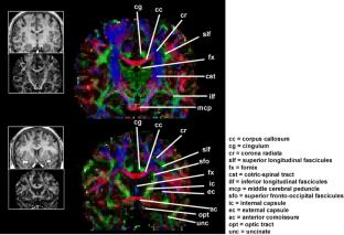
Diffusion tensor imaging (DTI) has become one of the most popular MRI techniques in brain research, as well as in clinical practice. The number of brain studies with DTI is growing steadily and, over the last decade, has produced more than 700 publications. Diffusion tensor imaging enables visualization and characterization of white matter fascicli in two and three dimensions. Since the introduction of this methodology in 1994, it has been used to study the white matter architecture and integrity of the normal and diseased brains (multiple sclerosis, stroke, aging, dementia, schizophrenia, etc.). Although it provided image contrast that was not available with routine MR techniques, unique information on white matter and 3D visualization of neuronal pathways, many questions were raised regarding the origin of the DTI signal. Diffusion tensor imaging is constantly validated, challenged, and developed in terms of acquisition scheme, image processing, analysis, and interpretation. While DTI offers a powerful tool to study and visualize white matter, it suffers from inherent artifacts and limitations. The partial volume effect and the inability of the model to cope with non-Gaussian diffusion are its two main drawbacks. Nevertheless, when combined with functional brain mapping, DTI provides an efficient tool for comprehensive, noninvasive, functional anatomy mapping of the human brain. This review summarizes the development of DTI in the last decade with respect to the specificity and utility of the technique in radiology and anatomy studies.
期刊介绍:
The Journal of Molecular Neuroscience is committed to the rapid publication of original findings that increase our understanding of the molecular structure, function, and development of the nervous system. The criteria for acceptance of manuscripts will be scientific excellence, originality, and relevance to the field of molecular neuroscience. Manuscripts with clinical relevance are especially encouraged since the journal seeks to provide a means for accelerating the progression of basic research findings toward clinical utilization. All experiments described in the Journal of Molecular Neuroscience that involve the use of animal or human subjects must have been approved by the appropriate institutional review committee and conform to accepted ethical standards.

 求助内容:
求助内容: 应助结果提醒方式:


