{"title":"评论:所有模型都是错误的,但有些是有用的:通过血管计算模型了解心房颤动的认知能力下降和痴呆","authors":"Brian Zenger, T. Jared Bunch","doi":"10.1002/ctd2.243","DOIUrl":null,"url":null,"abstract":"<p>Atrial fibrillation (AF) has been clearly shown to be an independent risk factor for cognitive decline and dementia.<span><sup>1, 2</sup></span> However, the exact mechanism that causes this precipitous cognitive decline is unknown.<span><sup>1</sup></span> Some of the most unmistakable evidence comes from the SWISS-AF trial, where adequately anticoagulated patients had serial brain imaging before and after starting anticoagulation therapy.<span><sup>3</sup></span> In this study, patients were shown to have an increased prevalence of brain injury compared to healthy controls that correlated with cognitive decline. A striking finding was that nearly all patients (99%) had white matter disease. The exact mechanisms for brain injury patterns beyond overt thrombotic or haemorrhagic mechanisms have been speculated to include many different hypotheses, including biochemical, drug side effects, vascular injury, or physiologic flow-related.<span><sup>1, 2, 4</sup></span> This region of brain vulnerability is supplied by the lenticulostriate arteries that have a perpendicular origin that is a potential vulnerable region to the haemodynamic perturbations of AF. Focal wall stress may result in vascular disease and dysfunction underlying what is traditionally described as “findings of chronic microvascular disease.” However, these mechanisms are hard to measure or test in humans and require advanced translational models for direct measurements. Computational modeling provides a unique avenue to test these hypotheses in a simulated setting with explicit parameter modulation based on patient-specific models.</p><p>In the most recent issue of the Journal of Clinical and Translational Medicine, Saglietto et al.<span><sup>5</sup></span> described their computational approach to showing how changes in wall shear stress in the lacunar brain vasculature during AF could contribute to the development of brain infarct outside of embolic events. The authors used well-established fluid dynamic simulation methods to show that during simulated episodes of AF, the wall shear stress range at the lacunar vessels junction is significantly increased compared to normal sinus rhythm. The authors show increased peak and trough wall shear stress localised to the vessel junctions. Importantly, changes in wall shear stress have been shown to have significant adverse downstream effects. In vitro data have shown that decreases in wall stress can lead to plaque buildup on the endothelium.<span><sup>6</sup></span> Furthermore, increased wall shear stress increases the likelihood of plaque rupture.<span><sup>6</sup></span> The authors postulate that increasing the dynamic range of sheer stress in the lacunar vessels during AF could create a vicious cycle of plaque buildup and rupture, leading to downstream brain infarction. Furthermore, the authors also highlight that increased wall stress alone can lead to long-term vessel damage, including atheroma formation, that could further develop into downstream infarction and microvascular dysfunction.<span><sup>6</sup></span></p><p>These results should not be surprising but quantify a previously handwavy point that the changes in flow or pressure in the brain vasculature during AF are meaningful to the development of downstream pathologies and dysfunction of adaptive physiology and autoregulation.<span><sup>7, 8</sup></span> Fundamentally, hypo/hyperperfusion events that occur during AF and cause changes in blood flow should drive changes in vascular sheer stress.<span><sup>7</sup></span> Until now, those suggestions have all been theoretical, with no firm grasp of the possible magnitude associated with AF episodes compared to normal sinus rhythm. This simulation study, which includes models from multiple patients, thousands of simulated heartbeats, and numerous anatomical differences, clearly demonstrates the range of changes that can occur during episodes of AF. Furthermore, we commend the authors’ creativity in linking in vitro data to simulation data to make clinically meaningful mechanistic statements.<span><sup>6</sup></span></p><p>A keen observer would note several flaws with simulated approaches, with this study no exception. Our response: all models are wrong; some models are useful. The assumptions in this study are multifactorial. The authors make many standard assumptions about vascular flow and changes in R–R intervals related to AF. They also assume a relatively static vessel with no differences associated with the dynamic flow in AF episodes. To our knowledge, these data are unavailable and, therefore, cannot be modelled. There are likely hyperlocal changes at these vascular junctions to reduce overall wall sheer stress. However, without highly invasive flow and pressure measurements, it would be impossible to determine those changes. Despite these fundamental assumptions, the downstream effect remains that patients with AF have a higher incidence of brain infarction and white matter injury than normal controls.<span><sup>3</sup></span> Therefore, these simulation results should not be considered invalid based on the assumptions discussed.</p><p>This study adds another hypothesis to the treasure trove of current hypotheses to relate AF to cognitive dysfunction and brain injury. As we already discussed, these hypotheses are broad from the molecular to the fluid dynamic level.<span><sup>2, 8</sup></span> Unfortunately, many studies focus exclusively on one measurement variable or parameter, including imaging, pressure, biochemical measurements, or other specific component. This exclusivity of studies makes it difficult to understand each variable's individual impact and contribution to the overall development of cognitive dysfunction. This makes it difficult to identify ideal clinical targets to reduce patient burden. The most likely scenario is a combination of these different hypotheses contributing to the development of various brain pathologies. Furthermore, it will be essential to understand how patient baseline characteristics affect these results and drive for more personalised care approaches (Figure 1).</p><p>Finally, this study adds more theoretical evidence for the value of early and aggressive rhythm control of AF. Recent clinical trials have shown a durable improvement in patient morbidity and mortality with effective early aggressive rhythm control and that AF ablation techniques outperform conventional medical management.<span><sup>9, 10</sup></span> These results, while not validated in a clinical trial, demonstrate the theoretical benefit that may be achieved with aggressive rhythm control compared to the medical management alone. Most clinical trials have focused on cardiac adverse events or stroke. These results suggest that cognitive dysfunction should also be considered a crucial secondary endpoint in patients suffering from AF.</p><p>The authors declare no conflict of interest.</p>","PeriodicalId":72605,"journal":{"name":"Clinical and translational discovery","volume":"3 5","pages":""},"PeriodicalIF":0.0000,"publicationDate":"2023-09-21","publicationTypes":"Journal Article","fieldsOfStudy":null,"isOpenAccess":false,"openAccessPdf":"https://onlinelibrary.wiley.com/doi/epdf/10.1002/ctd2.243","citationCount":"0","resultStr":"{\"title\":\"Commentary: All models are wrong, but some are useful: understanding cognitive decline and dementia in atrial fibrillation through vascular computational modelling\",\"authors\":\"Brian Zenger, T. Jared Bunch\",\"doi\":\"10.1002/ctd2.243\",\"DOIUrl\":null,\"url\":null,\"abstract\":\"<p>Atrial fibrillation (AF) has been clearly shown to be an independent risk factor for cognitive decline and dementia.<span><sup>1, 2</sup></span> However, the exact mechanism that causes this precipitous cognitive decline is unknown.<span><sup>1</sup></span> Some of the most unmistakable evidence comes from the SWISS-AF trial, where adequately anticoagulated patients had serial brain imaging before and after starting anticoagulation therapy.<span><sup>3</sup></span> In this study, patients were shown to have an increased prevalence of brain injury compared to healthy controls that correlated with cognitive decline. A striking finding was that nearly all patients (99%) had white matter disease. The exact mechanisms for brain injury patterns beyond overt thrombotic or haemorrhagic mechanisms have been speculated to include many different hypotheses, including biochemical, drug side effects, vascular injury, or physiologic flow-related.<span><sup>1, 2, 4</sup></span> This region of brain vulnerability is supplied by the lenticulostriate arteries that have a perpendicular origin that is a potential vulnerable region to the haemodynamic perturbations of AF. Focal wall stress may result in vascular disease and dysfunction underlying what is traditionally described as “findings of chronic microvascular disease.” However, these mechanisms are hard to measure or test in humans and require advanced translational models for direct measurements. Computational modeling provides a unique avenue to test these hypotheses in a simulated setting with explicit parameter modulation based on patient-specific models.</p><p>In the most recent issue of the Journal of Clinical and Translational Medicine, Saglietto et al.<span><sup>5</sup></span> described their computational approach to showing how changes in wall shear stress in the lacunar brain vasculature during AF could contribute to the development of brain infarct outside of embolic events. The authors used well-established fluid dynamic simulation methods to show that during simulated episodes of AF, the wall shear stress range at the lacunar vessels junction is significantly increased compared to normal sinus rhythm. The authors show increased peak and trough wall shear stress localised to the vessel junctions. Importantly, changes in wall shear stress have been shown to have significant adverse downstream effects. In vitro data have shown that decreases in wall stress can lead to plaque buildup on the endothelium.<span><sup>6</sup></span> Furthermore, increased wall shear stress increases the likelihood of plaque rupture.<span><sup>6</sup></span> The authors postulate that increasing the dynamic range of sheer stress in the lacunar vessels during AF could create a vicious cycle of plaque buildup and rupture, leading to downstream brain infarction. Furthermore, the authors also highlight that increased wall stress alone can lead to long-term vessel damage, including atheroma formation, that could further develop into downstream infarction and microvascular dysfunction.<span><sup>6</sup></span></p><p>These results should not be surprising but quantify a previously handwavy point that the changes in flow or pressure in the brain vasculature during AF are meaningful to the development of downstream pathologies and dysfunction of adaptive physiology and autoregulation.<span><sup>7, 8</sup></span> Fundamentally, hypo/hyperperfusion events that occur during AF and cause changes in blood flow should drive changes in vascular sheer stress.<span><sup>7</sup></span> Until now, those suggestions have all been theoretical, with no firm grasp of the possible magnitude associated with AF episodes compared to normal sinus rhythm. This simulation study, which includes models from multiple patients, thousands of simulated heartbeats, and numerous anatomical differences, clearly demonstrates the range of changes that can occur during episodes of AF. Furthermore, we commend the authors’ creativity in linking in vitro data to simulation data to make clinically meaningful mechanistic statements.<span><sup>6</sup></span></p><p>A keen observer would note several flaws with simulated approaches, with this study no exception. Our response: all models are wrong; some models are useful. The assumptions in this study are multifactorial. The authors make many standard assumptions about vascular flow and changes in R–R intervals related to AF. They also assume a relatively static vessel with no differences associated with the dynamic flow in AF episodes. To our knowledge, these data are unavailable and, therefore, cannot be modelled. There are likely hyperlocal changes at these vascular junctions to reduce overall wall sheer stress. However, without highly invasive flow and pressure measurements, it would be impossible to determine those changes. Despite these fundamental assumptions, the downstream effect remains that patients with AF have a higher incidence of brain infarction and white matter injury than normal controls.<span><sup>3</sup></span> Therefore, these simulation results should not be considered invalid based on the assumptions discussed.</p><p>This study adds another hypothesis to the treasure trove of current hypotheses to relate AF to cognitive dysfunction and brain injury. As we already discussed, these hypotheses are broad from the molecular to the fluid dynamic level.<span><sup>2, 8</sup></span> Unfortunately, many studies focus exclusively on one measurement variable or parameter, including imaging, pressure, biochemical measurements, or other specific component. This exclusivity of studies makes it difficult to understand each variable's individual impact and contribution to the overall development of cognitive dysfunction. This makes it difficult to identify ideal clinical targets to reduce patient burden. The most likely scenario is a combination of these different hypotheses contributing to the development of various brain pathologies. Furthermore, it will be essential to understand how patient baseline characteristics affect these results and drive for more personalised care approaches (Figure 1).</p><p>Finally, this study adds more theoretical evidence for the value of early and aggressive rhythm control of AF. Recent clinical trials have shown a durable improvement in patient morbidity and mortality with effective early aggressive rhythm control and that AF ablation techniques outperform conventional medical management.<span><sup>9, 10</sup></span> These results, while not validated in a clinical trial, demonstrate the theoretical benefit that may be achieved with aggressive rhythm control compared to the medical management alone. Most clinical trials have focused on cardiac adverse events or stroke. These results suggest that cognitive dysfunction should also be considered a crucial secondary endpoint in patients suffering from AF.</p><p>The authors declare no conflict of interest.</p>\",\"PeriodicalId\":72605,\"journal\":{\"name\":\"Clinical and translational discovery\",\"volume\":\"3 5\",\"pages\":\"\"},\"PeriodicalIF\":0.0000,\"publicationDate\":\"2023-09-21\",\"publicationTypes\":\"Journal Article\",\"fieldsOfStudy\":null,\"isOpenAccess\":false,\"openAccessPdf\":\"https://onlinelibrary.wiley.com/doi/epdf/10.1002/ctd2.243\",\"citationCount\":\"0\",\"resultStr\":null,\"platform\":\"Semanticscholar\",\"paperid\":null,\"PeriodicalName\":\"Clinical and translational discovery\",\"FirstCategoryId\":\"1085\",\"ListUrlMain\":\"https://onlinelibrary.wiley.com/doi/10.1002/ctd2.243\",\"RegionNum\":0,\"RegionCategory\":null,\"ArticlePicture\":[],\"TitleCN\":null,\"AbstractTextCN\":null,\"PMCID\":null,\"EPubDate\":\"\",\"PubModel\":\"\",\"JCR\":\"\",\"JCRName\":\"\",\"Score\":null,\"Total\":0}","platform":"Semanticscholar","paperid":null,"PeriodicalName":"Clinical and translational discovery","FirstCategoryId":"1085","ListUrlMain":"https://onlinelibrary.wiley.com/doi/10.1002/ctd2.243","RegionNum":0,"RegionCategory":null,"ArticlePicture":[],"TitleCN":null,"AbstractTextCN":null,"PMCID":null,"EPubDate":"","PubModel":"","JCR":"","JCRName":"","Score":null,"Total":0}
Commentary: All models are wrong, but some are useful: understanding cognitive decline and dementia in atrial fibrillation through vascular computational modelling
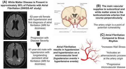
Atrial fibrillation (AF) has been clearly shown to be an independent risk factor for cognitive decline and dementia.1, 2 However, the exact mechanism that causes this precipitous cognitive decline is unknown.1 Some of the most unmistakable evidence comes from the SWISS-AF trial, where adequately anticoagulated patients had serial brain imaging before and after starting anticoagulation therapy.3 In this study, patients were shown to have an increased prevalence of brain injury compared to healthy controls that correlated with cognitive decline. A striking finding was that nearly all patients (99%) had white matter disease. The exact mechanisms for brain injury patterns beyond overt thrombotic or haemorrhagic mechanisms have been speculated to include many different hypotheses, including biochemical, drug side effects, vascular injury, or physiologic flow-related.1, 2, 4 This region of brain vulnerability is supplied by the lenticulostriate arteries that have a perpendicular origin that is a potential vulnerable region to the haemodynamic perturbations of AF. Focal wall stress may result in vascular disease and dysfunction underlying what is traditionally described as “findings of chronic microvascular disease.” However, these mechanisms are hard to measure or test in humans and require advanced translational models for direct measurements. Computational modeling provides a unique avenue to test these hypotheses in a simulated setting with explicit parameter modulation based on patient-specific models.
In the most recent issue of the Journal of Clinical and Translational Medicine, Saglietto et al.5 described their computational approach to showing how changes in wall shear stress in the lacunar brain vasculature during AF could contribute to the development of brain infarct outside of embolic events. The authors used well-established fluid dynamic simulation methods to show that during simulated episodes of AF, the wall shear stress range at the lacunar vessels junction is significantly increased compared to normal sinus rhythm. The authors show increased peak and trough wall shear stress localised to the vessel junctions. Importantly, changes in wall shear stress have been shown to have significant adverse downstream effects. In vitro data have shown that decreases in wall stress can lead to plaque buildup on the endothelium.6 Furthermore, increased wall shear stress increases the likelihood of plaque rupture.6 The authors postulate that increasing the dynamic range of sheer stress in the lacunar vessels during AF could create a vicious cycle of plaque buildup and rupture, leading to downstream brain infarction. Furthermore, the authors also highlight that increased wall stress alone can lead to long-term vessel damage, including atheroma formation, that could further develop into downstream infarction and microvascular dysfunction.6
These results should not be surprising but quantify a previously handwavy point that the changes in flow or pressure in the brain vasculature during AF are meaningful to the development of downstream pathologies and dysfunction of adaptive physiology and autoregulation.7, 8 Fundamentally, hypo/hyperperfusion events that occur during AF and cause changes in blood flow should drive changes in vascular sheer stress.7 Until now, those suggestions have all been theoretical, with no firm grasp of the possible magnitude associated with AF episodes compared to normal sinus rhythm. This simulation study, which includes models from multiple patients, thousands of simulated heartbeats, and numerous anatomical differences, clearly demonstrates the range of changes that can occur during episodes of AF. Furthermore, we commend the authors’ creativity in linking in vitro data to simulation data to make clinically meaningful mechanistic statements.6
A keen observer would note several flaws with simulated approaches, with this study no exception. Our response: all models are wrong; some models are useful. The assumptions in this study are multifactorial. The authors make many standard assumptions about vascular flow and changes in R–R intervals related to AF. They also assume a relatively static vessel with no differences associated with the dynamic flow in AF episodes. To our knowledge, these data are unavailable and, therefore, cannot be modelled. There are likely hyperlocal changes at these vascular junctions to reduce overall wall sheer stress. However, without highly invasive flow and pressure measurements, it would be impossible to determine those changes. Despite these fundamental assumptions, the downstream effect remains that patients with AF have a higher incidence of brain infarction and white matter injury than normal controls.3 Therefore, these simulation results should not be considered invalid based on the assumptions discussed.
This study adds another hypothesis to the treasure trove of current hypotheses to relate AF to cognitive dysfunction and brain injury. As we already discussed, these hypotheses are broad from the molecular to the fluid dynamic level.2, 8 Unfortunately, many studies focus exclusively on one measurement variable or parameter, including imaging, pressure, biochemical measurements, or other specific component. This exclusivity of studies makes it difficult to understand each variable's individual impact and contribution to the overall development of cognitive dysfunction. This makes it difficult to identify ideal clinical targets to reduce patient burden. The most likely scenario is a combination of these different hypotheses contributing to the development of various brain pathologies. Furthermore, it will be essential to understand how patient baseline characteristics affect these results and drive for more personalised care approaches (Figure 1).
Finally, this study adds more theoretical evidence for the value of early and aggressive rhythm control of AF. Recent clinical trials have shown a durable improvement in patient morbidity and mortality with effective early aggressive rhythm control and that AF ablation techniques outperform conventional medical management.9, 10 These results, while not validated in a clinical trial, demonstrate the theoretical benefit that may be achieved with aggressive rhythm control compared to the medical management alone. Most clinical trials have focused on cardiac adverse events or stroke. These results suggest that cognitive dysfunction should also be considered a crucial secondary endpoint in patients suffering from AF.

 求助内容:
求助内容: 应助结果提醒方式:


