{"title":"土耳其患者网状假核膜的多模式成像。","authors":"Serap Bilge Çeper, Filiz Afrashi, Cumali Değirmenci, Jale Menteş, Serhad Nalçacı, Cezmi Akkın","doi":"10.4274/tjo.galenos.2023.85616","DOIUrl":null,"url":null,"abstract":"<p><strong>Objectives: </strong>To investigate the presence and prevalence of reticular pseudodrusen (RPD) in patients with age-related macular degeneration using multiple imaging modalities and to compare the sensitivity and specificity of these modalities in the detection of RPD.</p><p><strong>Materials and methods: </strong>Images from a total of 198 consecutive patients were analyzed prospectively. Color fundus photography, red-free imaging, spectral domain optical coherence tomography (SD-OCT), infrared and blue reflectance (BR) imaging, fundus autofluorescence (FAF), enhanced-depth imaging OCT (EDI-OCT), fundus fluorescein angiography (FFA) and indocyanine green angiography were performed. RPD was diagnosed in the presence of relevant findings in at least two of the imaging methods used.</p><p><strong>Results: </strong>RPD were detected in 149 eyes (37.6%). In the detection of RPD, color fundus photography, red-free photography, SD-OCT, infrared, FAF, BR, and FFA imaging had sensitivity values of 50%, 57.7%, 91.6%, 95%, 74.6%, 65.7%, and 28.2% and specificity values of 99.6%, 100%, 98.4%, 94.6%, 100%, 99.6%, and 69.8%, respectively.</p><p><strong>Conclusion: </strong>Infrared imaging had the highest sensitivity. SD-OCT combined with infrared imaging was the most sensitive imaging technique for detecting RPD. The high specificity of FAF, red-free, and BR imaging may be useful to confirm a diagnosis of RPD.</p>","PeriodicalId":23373,"journal":{"name":"Turkish Journal of Ophthalmology","volume":"53 5","pages":"275-280"},"PeriodicalIF":0.0000,"publicationDate":"2023-10-19","publicationTypes":"Journal Article","fieldsOfStudy":null,"isOpenAccess":false,"openAccessPdf":"https://ftp.ncbi.nlm.nih.gov/pub/pmc/oa_pdf/ba/4e/TJO-53-275.PMC10599339.pdf","citationCount":"0","resultStr":"{\"title\":\"Multimodal Imaging of Reticular Pseudodrusen in Turkish Patients.\",\"authors\":\"Serap Bilge Çeper, Filiz Afrashi, Cumali Değirmenci, Jale Menteş, Serhad Nalçacı, Cezmi Akkın\",\"doi\":\"10.4274/tjo.galenos.2023.85616\",\"DOIUrl\":null,\"url\":null,\"abstract\":\"<p><strong>Objectives: </strong>To investigate the presence and prevalence of reticular pseudodrusen (RPD) in patients with age-related macular degeneration using multiple imaging modalities and to compare the sensitivity and specificity of these modalities in the detection of RPD.</p><p><strong>Materials and methods: </strong>Images from a total of 198 consecutive patients were analyzed prospectively. Color fundus photography, red-free imaging, spectral domain optical coherence tomography (SD-OCT), infrared and blue reflectance (BR) imaging, fundus autofluorescence (FAF), enhanced-depth imaging OCT (EDI-OCT), fundus fluorescein angiography (FFA) and indocyanine green angiography were performed. RPD was diagnosed in the presence of relevant findings in at least two of the imaging methods used.</p><p><strong>Results: </strong>RPD were detected in 149 eyes (37.6%). In the detection of RPD, color fundus photography, red-free photography, SD-OCT, infrared, FAF, BR, and FFA imaging had sensitivity values of 50%, 57.7%, 91.6%, 95%, 74.6%, 65.7%, and 28.2% and specificity values of 99.6%, 100%, 98.4%, 94.6%, 100%, 99.6%, and 69.8%, respectively.</p><p><strong>Conclusion: </strong>Infrared imaging had the highest sensitivity. SD-OCT combined with infrared imaging was the most sensitive imaging technique for detecting RPD. The high specificity of FAF, red-free, and BR imaging may be useful to confirm a diagnosis of RPD.</p>\",\"PeriodicalId\":23373,\"journal\":{\"name\":\"Turkish Journal of Ophthalmology\",\"volume\":\"53 5\",\"pages\":\"275-280\"},\"PeriodicalIF\":0.0000,\"publicationDate\":\"2023-10-19\",\"publicationTypes\":\"Journal Article\",\"fieldsOfStudy\":null,\"isOpenAccess\":false,\"openAccessPdf\":\"https://ftp.ncbi.nlm.nih.gov/pub/pmc/oa_pdf/ba/4e/TJO-53-275.PMC10599339.pdf\",\"citationCount\":\"0\",\"resultStr\":null,\"platform\":\"Semanticscholar\",\"paperid\":null,\"PeriodicalName\":\"Turkish Journal of Ophthalmology\",\"FirstCategoryId\":\"1085\",\"ListUrlMain\":\"https://doi.org/10.4274/tjo.galenos.2023.85616\",\"RegionNum\":0,\"RegionCategory\":null,\"ArticlePicture\":[],\"TitleCN\":null,\"AbstractTextCN\":null,\"PMCID\":null,\"EPubDate\":\"\",\"PubModel\":\"\",\"JCR\":\"Q3\",\"JCRName\":\"Medicine\",\"Score\":null,\"Total\":0}","platform":"Semanticscholar","paperid":null,"PeriodicalName":"Turkish Journal of Ophthalmology","FirstCategoryId":"1085","ListUrlMain":"https://doi.org/10.4274/tjo.galenos.2023.85616","RegionNum":0,"RegionCategory":null,"ArticlePicture":[],"TitleCN":null,"AbstractTextCN":null,"PMCID":null,"EPubDate":"","PubModel":"","JCR":"Q3","JCRName":"Medicine","Score":null,"Total":0}
Multimodal Imaging of Reticular Pseudodrusen in Turkish Patients.
Objectives: To investigate the presence and prevalence of reticular pseudodrusen (RPD) in patients with age-related macular degeneration using multiple imaging modalities and to compare the sensitivity and specificity of these modalities in the detection of RPD.
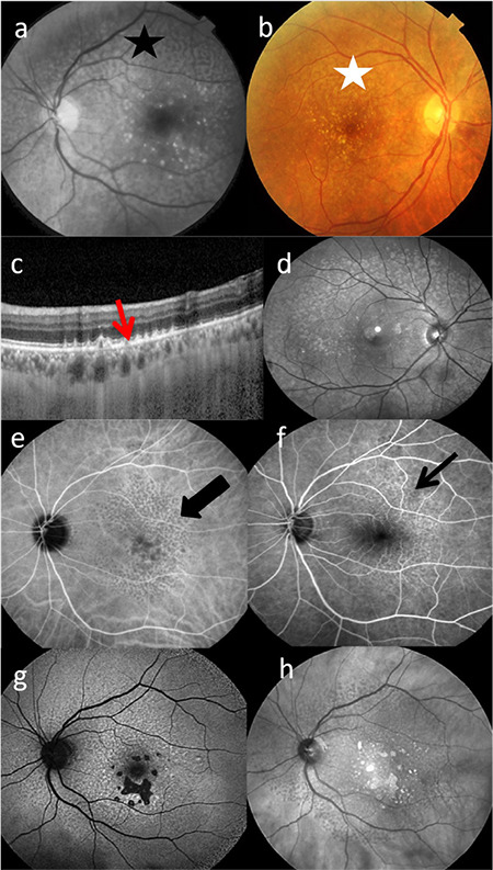
Materials and methods: Images from a total of 198 consecutive patients were analyzed prospectively. Color fundus photography, red-free imaging, spectral domain optical coherence tomography (SD-OCT), infrared and blue reflectance (BR) imaging, fundus autofluorescence (FAF), enhanced-depth imaging OCT (EDI-OCT), fundus fluorescein angiography (FFA) and indocyanine green angiography were performed. RPD was diagnosed in the presence of relevant findings in at least two of the imaging methods used.
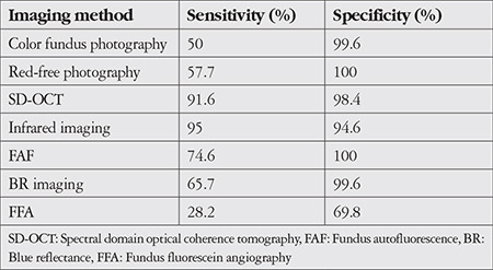
Results: RPD were detected in 149 eyes (37.6%). In the detection of RPD, color fundus photography, red-free photography, SD-OCT, infrared, FAF, BR, and FFA imaging had sensitivity values of 50%, 57.7%, 91.6%, 95%, 74.6%, 65.7%, and 28.2% and specificity values of 99.6%, 100%, 98.4%, 94.6%, 100%, 99.6%, and 69.8%, respectively.
Conclusion: Infrared imaging had the highest sensitivity. SD-OCT combined with infrared imaging was the most sensitive imaging technique for detecting RPD. The high specificity of FAF, red-free, and BR imaging may be useful to confirm a diagnosis of RPD.
期刊介绍:
The Turkish Journal of Ophthalmology (TJO) is the only scientific periodical publication of the Turkish Ophthalmological Association and has been published since January 1929. In its early years, the journal was published in Turkish and French. Although there were temporary interruptions in the publication of the journal due to various challenges, the Turkish Journal of Ophthalmology has been published continually from 1971 to the present. The target audience includes specialists and physicians in training in ophthalmology in all relevant disciplines.

 求助内容:
求助内容: 应助结果提醒方式:


