{"title":"使用基于桌面的无代码机器学习工具检测和分类糖尿病黄斑水肿。","authors":"Furkan Kırık, Büşra Demirkıran, Cansu Ekinci Aslanoğlu, Arif Koytak, Hakan Özdemir","doi":"10.4274/tjo.galenos.2023.92635","DOIUrl":null,"url":null,"abstract":"<p><strong>Objectives: </strong>To evaluate the effectiveness of the Lobe application, a machine learning (ML) tool that can be used on a personal computer without requiring coding expertise, in the recognition and classification of diabetic macular edema (DME) in spectral-domain optical coherence tomography (SD-OCT) scans.</p><p><strong>Materials and methods: </strong>A total of 695 cross-sectional SD-OCT images from 336 patients with DME and 200 OCT images of 200 healthy controls were included. Images with DME were classified into three main types: diffuse retinal edema (DRE), cystoid macular edema (CME), and cystoid macular degeneration (CMD). To develop the ML model, we used the desktop-based code-free Lobe application, which includes a pre-trained ResNet-50 V2 convolutional neural network and is available free of charge. The performance of the trained model in recognizing and classifying DME was evaluated with 41 DRE, 28 CMD, 70 CME, and 40 normal SD-OCT images that were not used in the training.</p><p><strong>Results: </strong>The developed model showed 99.28% sensitivity and 100% specificity for class-independent detection of DME. Sensitivity and specificity by labels were 87.80% and 98.57% for DRE, 96.43% and 99.29% for CME, and 95.71% and 95.41% for CMD, respectively.</p><p><strong>Conclusion: </strong>To our knowledge, this is the first evaluation of the effectiveness of Lobe with ophthalmological images, and the results indicate that it can be used with high efficiency in the recognition and classification of DME from SD-OCT images by ophthalmologists without coding expertise.</p>","PeriodicalId":23373,"journal":{"name":"Turkish Journal of Ophthalmology","volume":"53 5","pages":"301-306"},"PeriodicalIF":0.0000,"publicationDate":"2023-10-19","publicationTypes":"Journal Article","fieldsOfStudy":null,"isOpenAccess":false,"openAccessPdf":"https://ftp.ncbi.nlm.nih.gov/pub/pmc/oa_pdf/a1/da/TJO-53-301.PMC10599341.pdf","citationCount":"0","resultStr":"{\"title\":\"Detection and Classification of Diabetic Macular Edema with a Desktop-Based Code-Free Machine Learning Tool.\",\"authors\":\"Furkan Kırık, Büşra Demirkıran, Cansu Ekinci Aslanoğlu, Arif Koytak, Hakan Özdemir\",\"doi\":\"10.4274/tjo.galenos.2023.92635\",\"DOIUrl\":null,\"url\":null,\"abstract\":\"<p><strong>Objectives: </strong>To evaluate the effectiveness of the Lobe application, a machine learning (ML) tool that can be used on a personal computer without requiring coding expertise, in the recognition and classification of diabetic macular edema (DME) in spectral-domain optical coherence tomography (SD-OCT) scans.</p><p><strong>Materials and methods: </strong>A total of 695 cross-sectional SD-OCT images from 336 patients with DME and 200 OCT images of 200 healthy controls were included. Images with DME were classified into three main types: diffuse retinal edema (DRE), cystoid macular edema (CME), and cystoid macular degeneration (CMD). To develop the ML model, we used the desktop-based code-free Lobe application, which includes a pre-trained ResNet-50 V2 convolutional neural network and is available free of charge. The performance of the trained model in recognizing and classifying DME was evaluated with 41 DRE, 28 CMD, 70 CME, and 40 normal SD-OCT images that were not used in the training.</p><p><strong>Results: </strong>The developed model showed 99.28% sensitivity and 100% specificity for class-independent detection of DME. Sensitivity and specificity by labels were 87.80% and 98.57% for DRE, 96.43% and 99.29% for CME, and 95.71% and 95.41% for CMD, respectively.</p><p><strong>Conclusion: </strong>To our knowledge, this is the first evaluation of the effectiveness of Lobe with ophthalmological images, and the results indicate that it can be used with high efficiency in the recognition and classification of DME from SD-OCT images by ophthalmologists without coding expertise.</p>\",\"PeriodicalId\":23373,\"journal\":{\"name\":\"Turkish Journal of Ophthalmology\",\"volume\":\"53 5\",\"pages\":\"301-306\"},\"PeriodicalIF\":0.0000,\"publicationDate\":\"2023-10-19\",\"publicationTypes\":\"Journal Article\",\"fieldsOfStudy\":null,\"isOpenAccess\":false,\"openAccessPdf\":\"https://ftp.ncbi.nlm.nih.gov/pub/pmc/oa_pdf/a1/da/TJO-53-301.PMC10599341.pdf\",\"citationCount\":\"0\",\"resultStr\":null,\"platform\":\"Semanticscholar\",\"paperid\":null,\"PeriodicalName\":\"Turkish Journal of Ophthalmology\",\"FirstCategoryId\":\"1085\",\"ListUrlMain\":\"https://doi.org/10.4274/tjo.galenos.2023.92635\",\"RegionNum\":0,\"RegionCategory\":null,\"ArticlePicture\":[],\"TitleCN\":null,\"AbstractTextCN\":null,\"PMCID\":null,\"EPubDate\":\"\",\"PubModel\":\"\",\"JCR\":\"Q3\",\"JCRName\":\"Medicine\",\"Score\":null,\"Total\":0}","platform":"Semanticscholar","paperid":null,"PeriodicalName":"Turkish Journal of Ophthalmology","FirstCategoryId":"1085","ListUrlMain":"https://doi.org/10.4274/tjo.galenos.2023.92635","RegionNum":0,"RegionCategory":null,"ArticlePicture":[],"TitleCN":null,"AbstractTextCN":null,"PMCID":null,"EPubDate":"","PubModel":"","JCR":"Q3","JCRName":"Medicine","Score":null,"Total":0}
Detection and Classification of Diabetic Macular Edema with a Desktop-Based Code-Free Machine Learning Tool.
Objectives: To evaluate the effectiveness of the Lobe application, a machine learning (ML) tool that can be used on a personal computer without requiring coding expertise, in the recognition and classification of diabetic macular edema (DME) in spectral-domain optical coherence tomography (SD-OCT) scans.
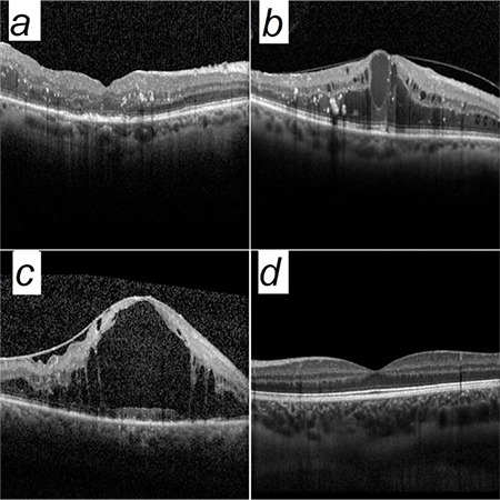
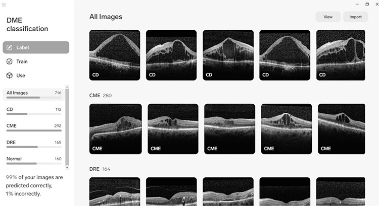
Materials and methods: A total of 695 cross-sectional SD-OCT images from 336 patients with DME and 200 OCT images of 200 healthy controls were included. Images with DME were classified into three main types: diffuse retinal edema (DRE), cystoid macular edema (CME), and cystoid macular degeneration (CMD). To develop the ML model, we used the desktop-based code-free Lobe application, which includes a pre-trained ResNet-50 V2 convolutional neural network and is available free of charge. The performance of the trained model in recognizing and classifying DME was evaluated with 41 DRE, 28 CMD, 70 CME, and 40 normal SD-OCT images that were not used in the training.
Results: The developed model showed 99.28% sensitivity and 100% specificity for class-independent detection of DME. Sensitivity and specificity by labels were 87.80% and 98.57% for DRE, 96.43% and 99.29% for CME, and 95.71% and 95.41% for CMD, respectively.
Conclusion: To our knowledge, this is the first evaluation of the effectiveness of Lobe with ophthalmological images, and the results indicate that it can be used with high efficiency in the recognition and classification of DME from SD-OCT images by ophthalmologists without coding expertise.
期刊介绍:
The Turkish Journal of Ophthalmology (TJO) is the only scientific periodical publication of the Turkish Ophthalmological Association and has been published since January 1929. In its early years, the journal was published in Turkish and French. Although there were temporary interruptions in the publication of the journal due to various challenges, the Turkish Journal of Ophthalmology has been published continually from 1971 to the present. The target audience includes specialists and physicians in training in ophthalmology in all relevant disciplines.


 求助内容:
求助内容: 应助结果提醒方式:


