Ayşin Tuba Kaplan, Sibel Öskan Yalçın, Safiye Güneş Sağer
{"title":"小儿视乳头水肿和假性视乳头水肿的临床表现和光学相干断层扫描测量。","authors":"Ayşin Tuba Kaplan, Sibel Öskan Yalçın, Safiye Güneş Sağer","doi":"10.4274/tjo.galenos.2023.81504","DOIUrl":null,"url":null,"abstract":"<p><strong>Objectives: </strong>To compare the clinical findings and multimodal imaging of pediatric patients diagnosed with papilledema and pseudopapilledema with those of healthy individuals.</p><p><strong>Materials and methods: </strong>Ninety children (<18 years of age) referred for suspected papilledema were included in this study. All patients underwent optical coherence tomography (OCT) imaging and were compared with normal control subjects.</p><p><strong>Results: </strong>Fifty-eight children diagnosed with pseudopapilledema, 32 children with mild-to-moderate papilledema, and 40 controls were evaluated. The average and all quadrants of retinal nerve fiber layer (RNFL) thickness were significantly higher in the papilledema group than in the pseudopapilledema and control groups (p<0.001). Bruch's membrane opening (BMO) measurements were similar in both groups (p>0.05). The average, nasal, and temporal RNFL thicknesses were significantly higher in the pseudopapilledema group compared with the controls (p<0.001). Area under the receiver operating characteristic (ROC) curve showed high diagnostic ability for RNFL thickness in all quadrants to differentiate papilledema from pseudopapilledema (p<0.001). In the pseudopapilledema group, average, temporal, and inferior RNFL thickness and BMO measurements were significantly higher in eyes with optic nerve head drusen (n=28) compared with those without drusen (n=88) (p=0.035, p=0.022, p=0.040 and, p=0.047 respectively).</p><p><strong>Conclusion: </strong>Papilledema and pseudopapilledema show great differences in evaluation, follow-up, and prognosis. Using non-invasive methods such as newly developed OCT techniques in differential diagnosis can relieve patients with pseudopapilledema from the stress and financial burden of expensive, extensive, and invasive procedures.</p>","PeriodicalId":23373,"journal":{"name":"Turkish Journal of Ophthalmology","volume":"53 5","pages":"294-300"},"PeriodicalIF":0.0000,"publicationDate":"2023-10-19","publicationTypes":"Journal Article","fieldsOfStudy":null,"isOpenAccess":false,"openAccessPdf":"https://ftp.ncbi.nlm.nih.gov/pub/pmc/oa_pdf/14/d6/TJO-53-294.PMC10599342.pdf","citationCount":"0","resultStr":"{\"title\":\"Clinical Findings and Optical Coherence Tomography Measurements of Pediatric Patients with Papilledema and Pseudopapilledema.\",\"authors\":\"Ayşin Tuba Kaplan, Sibel Öskan Yalçın, Safiye Güneş Sağer\",\"doi\":\"10.4274/tjo.galenos.2023.81504\",\"DOIUrl\":null,\"url\":null,\"abstract\":\"<p><strong>Objectives: </strong>To compare the clinical findings and multimodal imaging of pediatric patients diagnosed with papilledema and pseudopapilledema with those of healthy individuals.</p><p><strong>Materials and methods: </strong>Ninety children (<18 years of age) referred for suspected papilledema were included in this study. All patients underwent optical coherence tomography (OCT) imaging and were compared with normal control subjects.</p><p><strong>Results: </strong>Fifty-eight children diagnosed with pseudopapilledema, 32 children with mild-to-moderate papilledema, and 40 controls were evaluated. The average and all quadrants of retinal nerve fiber layer (RNFL) thickness were significantly higher in the papilledema group than in the pseudopapilledema and control groups (p<0.001). Bruch's membrane opening (BMO) measurements were similar in both groups (p>0.05). The average, nasal, and temporal RNFL thicknesses were significantly higher in the pseudopapilledema group compared with the controls (p<0.001). Area under the receiver operating characteristic (ROC) curve showed high diagnostic ability for RNFL thickness in all quadrants to differentiate papilledema from pseudopapilledema (p<0.001). In the pseudopapilledema group, average, temporal, and inferior RNFL thickness and BMO measurements were significantly higher in eyes with optic nerve head drusen (n=28) compared with those without drusen (n=88) (p=0.035, p=0.022, p=0.040 and, p=0.047 respectively).</p><p><strong>Conclusion: </strong>Papilledema and pseudopapilledema show great differences in evaluation, follow-up, and prognosis. Using non-invasive methods such as newly developed OCT techniques in differential diagnosis can relieve patients with pseudopapilledema from the stress and financial burden of expensive, extensive, and invasive procedures.</p>\",\"PeriodicalId\":23373,\"journal\":{\"name\":\"Turkish Journal of Ophthalmology\",\"volume\":\"53 5\",\"pages\":\"294-300\"},\"PeriodicalIF\":0.0000,\"publicationDate\":\"2023-10-19\",\"publicationTypes\":\"Journal Article\",\"fieldsOfStudy\":null,\"isOpenAccess\":false,\"openAccessPdf\":\"https://ftp.ncbi.nlm.nih.gov/pub/pmc/oa_pdf/14/d6/TJO-53-294.PMC10599342.pdf\",\"citationCount\":\"0\",\"resultStr\":null,\"platform\":\"Semanticscholar\",\"paperid\":null,\"PeriodicalName\":\"Turkish Journal of Ophthalmology\",\"FirstCategoryId\":\"1085\",\"ListUrlMain\":\"https://doi.org/10.4274/tjo.galenos.2023.81504\",\"RegionNum\":0,\"RegionCategory\":null,\"ArticlePicture\":[],\"TitleCN\":null,\"AbstractTextCN\":null,\"PMCID\":null,\"EPubDate\":\"\",\"PubModel\":\"\",\"JCR\":\"Q3\",\"JCRName\":\"Medicine\",\"Score\":null,\"Total\":0}","platform":"Semanticscholar","paperid":null,"PeriodicalName":"Turkish Journal of Ophthalmology","FirstCategoryId":"1085","ListUrlMain":"https://doi.org/10.4274/tjo.galenos.2023.81504","RegionNum":0,"RegionCategory":null,"ArticlePicture":[],"TitleCN":null,"AbstractTextCN":null,"PMCID":null,"EPubDate":"","PubModel":"","JCR":"Q3","JCRName":"Medicine","Score":null,"Total":0}
Clinical Findings and Optical Coherence Tomography Measurements of Pediatric Patients with Papilledema and Pseudopapilledema.
Objectives: To compare the clinical findings and multimodal imaging of pediatric patients diagnosed with papilledema and pseudopapilledema with those of healthy individuals.
Materials and methods: Ninety children (<18 years of age) referred for suspected papilledema were included in this study. All patients underwent optical coherence tomography (OCT) imaging and were compared with normal control subjects.
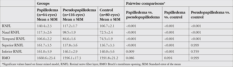
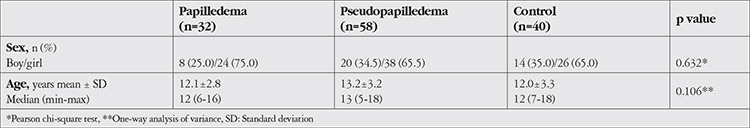
Results: Fifty-eight children diagnosed with pseudopapilledema, 32 children with mild-to-moderate papilledema, and 40 controls were evaluated. The average and all quadrants of retinal nerve fiber layer (RNFL) thickness were significantly higher in the papilledema group than in the pseudopapilledema and control groups (p<0.001). Bruch's membrane opening (BMO) measurements were similar in both groups (p>0.05). The average, nasal, and temporal RNFL thicknesses were significantly higher in the pseudopapilledema group compared with the controls (p<0.001). Area under the receiver operating characteristic (ROC) curve showed high diagnostic ability for RNFL thickness in all quadrants to differentiate papilledema from pseudopapilledema (p<0.001). In the pseudopapilledema group, average, temporal, and inferior RNFL thickness and BMO measurements were significantly higher in eyes with optic nerve head drusen (n=28) compared with those without drusen (n=88) (p=0.035, p=0.022, p=0.040 and, p=0.047 respectively).
Conclusion: Papilledema and pseudopapilledema show great differences in evaluation, follow-up, and prognosis. Using non-invasive methods such as newly developed OCT techniques in differential diagnosis can relieve patients with pseudopapilledema from the stress and financial burden of expensive, extensive, and invasive procedures.
期刊介绍:
The Turkish Journal of Ophthalmology (TJO) is the only scientific periodical publication of the Turkish Ophthalmological Association and has been published since January 1929. In its early years, the journal was published in Turkish and French. Although there were temporary interruptions in the publication of the journal due to various challenges, the Turkish Journal of Ophthalmology has been published continually from 1971 to the present. The target audience includes specialists and physicians in training in ophthalmology in all relevant disciplines.

 求助内容:
求助内容: 应助结果提醒方式:


