Eva Aalbregt, Lotte Rijken, Aart Nederveen, Pim van Ooij, Kak Khee Yeung, Vincent Jongkind
{"title":"定量磁共振成像评估主动脉瘤的进展和破裂风险:范围界定综述。","authors":"Eva Aalbregt, Lotte Rijken, Aart Nederveen, Pim van Ooij, Kak Khee Yeung, Vincent Jongkind","doi":"10.1177/15266028231204830","DOIUrl":null,"url":null,"abstract":"<p><strong>Purpose: </strong>In current practice, the diameter of an aortic aneurysm is utilized to estimate the rupture risk and decide upon timing of elective repair, although it is known to be imprecise and not patient-specific. Quantitative magnetic resonance imaging (MRI) enables the visualization of several biomarkers that provide information about processes within the aneurysm and may therefore facilitate patient-specific risk stratification. We performed a scoping review of the literature on quantitative MRI techniques to assess aortic aneurysm progression and rupture risk, summarized these findings, and identified knowledge gaps.</p><p><strong>Methods: </strong>Literature concerning primary research was of interest and the medical databases PubMed, Scopus, Embase, and Cochrane were systematically searched. This study used the PRISMA protocol extension for scoping reviews. Articles published between January 2010 and February 2023 involving animals and/or humans were included. Data were extracted by 2 authors using a predefined charting method.</p><p><strong>Results: </strong>A total of 1641 articles were identified, of which 21 were included in the scoping review. Quantitative MRI-derived biomarkers were categorized into hemodynamic (8 studies), wall (5 studies) and molecular biomarkers (8 studies). Fifteen studies included patients and/or healthy human subjects. Animal models were investigated in the other 6 studies. A cross-sectional study design was the most common, whereas 5 animal studies had a longitudinal component and 2 studies including patients had a prospective design. A promising hemodynamic biomarker is wall shear stress (WSS), which is estimated based on 4D-flow MRI. Molecular biomarkers enable the assessment of inflammatory and wall deterioration processes. The ADAMTS4-specific molecular magnetic resonance (MR) probe showed potential to predict abdominal aortic aneurysm (AAA) formation and rupture in a murine model. Wall biomarkers assessed using dynamic contrast-enhanced (DCE) MRI showed great potential for assessing AAA progression independent of the maximum diameter.</p><p><strong>Conclusion: </strong>This scoping review provides an overview of quantitative MRI techniques studied and the biomarkers derived from them to assess aortic aneurysm progression and rupture risk. Longitudinal studies are needed to validate the causal relationships between the identified biomarkers and aneurysm growth, rupture, or repair. In the future, quantitative MRI could play an important role in the personalized risk assessment of aortic aneurysm rupture.Clinical ImpactThe currently used maximum aneurysm diameter fails to accurately assess the multifactorial pathology of an aortic aneurysm and precisely predicts rupture in a patient-specific manner. Quantitative magnetic resonance imaging (MRI) enables the detection of various quantitative parameters involved in aneurysm progression and subsequent rupture. This scoping review provides an overview of the studied quantitative MRI techniques, the biomarkers derived from them, and recommendations for future research needed for the implementation of these biomarkers. Ultimately, quantitative MRI could facilitate personalized risk assessment for patients with aortic aneurysms, thereby reducing untimely repairs and improving rupture prevention.</p>","PeriodicalId":50210,"journal":{"name":"Journal of Endovascular Therapy","volume":" ","pages":"929-945"},"PeriodicalIF":1.5000,"publicationDate":"2025-08-01","publicationTypes":"Journal Article","fieldsOfStudy":null,"isOpenAccess":false,"openAccessPdf":"https://www.ncbi.nlm.nih.gov/pmc/articles/PMC12241695/pdf/","citationCount":"0","resultStr":"{\"title\":\"Quantitative Magnetic Resonance Imaging to Assess Progression and Rupture Risk of Aortic Aneurysms: A Scoping Review.\",\"authors\":\"Eva Aalbregt, Lotte Rijken, Aart Nederveen, Pim van Ooij, Kak Khee Yeung, Vincent Jongkind\",\"doi\":\"10.1177/15266028231204830\",\"DOIUrl\":null,\"url\":null,\"abstract\":\"<p><strong>Purpose: </strong>In current practice, the diameter of an aortic aneurysm is utilized to estimate the rupture risk and decide upon timing of elective repair, although it is known to be imprecise and not patient-specific. Quantitative magnetic resonance imaging (MRI) enables the visualization of several biomarkers that provide information about processes within the aneurysm and may therefore facilitate patient-specific risk stratification. We performed a scoping review of the literature on quantitative MRI techniques to assess aortic aneurysm progression and rupture risk, summarized these findings, and identified knowledge gaps.</p><p><strong>Methods: </strong>Literature concerning primary research was of interest and the medical databases PubMed, Scopus, Embase, and Cochrane were systematically searched. This study used the PRISMA protocol extension for scoping reviews. Articles published between January 2010 and February 2023 involving animals and/or humans were included. Data were extracted by 2 authors using a predefined charting method.</p><p><strong>Results: </strong>A total of 1641 articles were identified, of which 21 were included in the scoping review. Quantitative MRI-derived biomarkers were categorized into hemodynamic (8 studies), wall (5 studies) and molecular biomarkers (8 studies). Fifteen studies included patients and/or healthy human subjects. Animal models were investigated in the other 6 studies. A cross-sectional study design was the most common, whereas 5 animal studies had a longitudinal component and 2 studies including patients had a prospective design. A promising hemodynamic biomarker is wall shear stress (WSS), which is estimated based on 4D-flow MRI. Molecular biomarkers enable the assessment of inflammatory and wall deterioration processes. The ADAMTS4-specific molecular magnetic resonance (MR) probe showed potential to predict abdominal aortic aneurysm (AAA) formation and rupture in a murine model. Wall biomarkers assessed using dynamic contrast-enhanced (DCE) MRI showed great potential for assessing AAA progression independent of the maximum diameter.</p><p><strong>Conclusion: </strong>This scoping review provides an overview of quantitative MRI techniques studied and the biomarkers derived from them to assess aortic aneurysm progression and rupture risk. Longitudinal studies are needed to validate the causal relationships between the identified biomarkers and aneurysm growth, rupture, or repair. In the future, quantitative MRI could play an important role in the personalized risk assessment of aortic aneurysm rupture.Clinical ImpactThe currently used maximum aneurysm diameter fails to accurately assess the multifactorial pathology of an aortic aneurysm and precisely predicts rupture in a patient-specific manner. Quantitative magnetic resonance imaging (MRI) enables the detection of various quantitative parameters involved in aneurysm progression and subsequent rupture. This scoping review provides an overview of the studied quantitative MRI techniques, the biomarkers derived from them, and recommendations for future research needed for the implementation of these biomarkers. Ultimately, quantitative MRI could facilitate personalized risk assessment for patients with aortic aneurysms, thereby reducing untimely repairs and improving rupture prevention.</p>\",\"PeriodicalId\":50210,\"journal\":{\"name\":\"Journal of Endovascular Therapy\",\"volume\":\" \",\"pages\":\"929-945\"},\"PeriodicalIF\":1.5000,\"publicationDate\":\"2025-08-01\",\"publicationTypes\":\"Journal Article\",\"fieldsOfStudy\":null,\"isOpenAccess\":false,\"openAccessPdf\":\"https://www.ncbi.nlm.nih.gov/pmc/articles/PMC12241695/pdf/\",\"citationCount\":\"0\",\"resultStr\":null,\"platform\":\"Semanticscholar\",\"paperid\":null,\"PeriodicalName\":\"Journal of Endovascular Therapy\",\"FirstCategoryId\":\"3\",\"ListUrlMain\":\"https://doi.org/10.1177/15266028231204830\",\"RegionNum\":2,\"RegionCategory\":\"医学\",\"ArticlePicture\":[],\"TitleCN\":null,\"AbstractTextCN\":null,\"PMCID\":null,\"EPubDate\":\"2023/10/18 0:00:00\",\"PubModel\":\"Epub\",\"JCR\":\"Q3\",\"JCRName\":\"PERIPHERAL VASCULAR DISEASE\",\"Score\":null,\"Total\":0}","platform":"Semanticscholar","paperid":null,"PeriodicalName":"Journal of Endovascular Therapy","FirstCategoryId":"3","ListUrlMain":"https://doi.org/10.1177/15266028231204830","RegionNum":2,"RegionCategory":"医学","ArticlePicture":[],"TitleCN":null,"AbstractTextCN":null,"PMCID":null,"EPubDate":"2023/10/18 0:00:00","PubModel":"Epub","JCR":"Q3","JCRName":"PERIPHERAL VASCULAR DISEASE","Score":null,"Total":0}
Quantitative Magnetic Resonance Imaging to Assess Progression and Rupture Risk of Aortic Aneurysms: A Scoping Review.
Purpose: In current practice, the diameter of an aortic aneurysm is utilized to estimate the rupture risk and decide upon timing of elective repair, although it is known to be imprecise and not patient-specific. Quantitative magnetic resonance imaging (MRI) enables the visualization of several biomarkers that provide information about processes within the aneurysm and may therefore facilitate patient-specific risk stratification. We performed a scoping review of the literature on quantitative MRI techniques to assess aortic aneurysm progression and rupture risk, summarized these findings, and identified knowledge gaps.
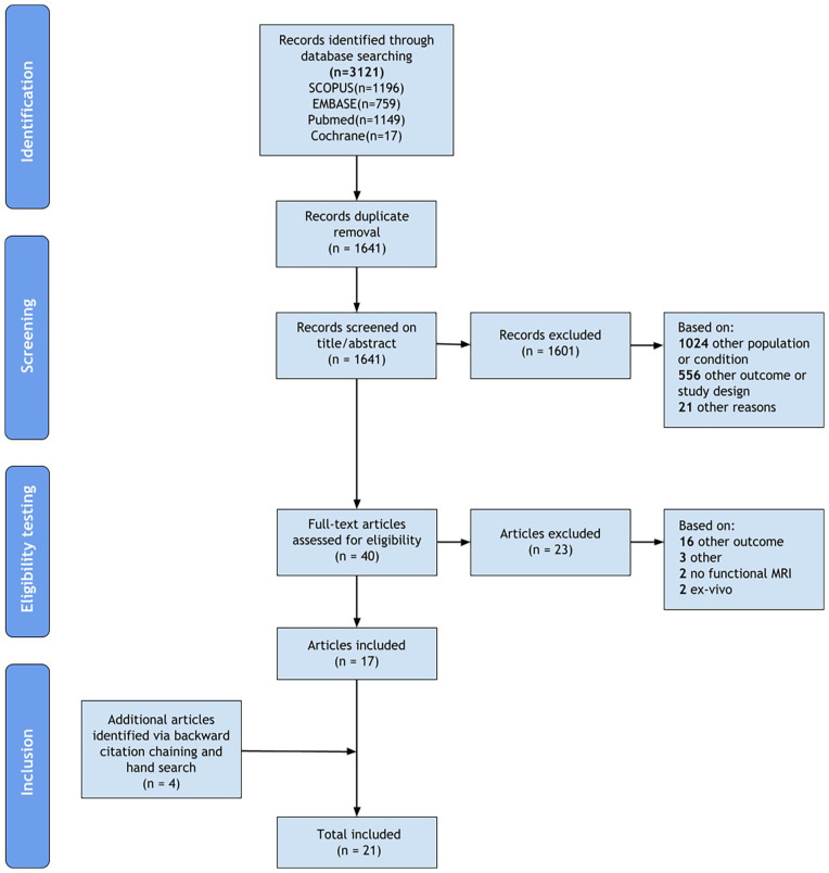
Methods: Literature concerning primary research was of interest and the medical databases PubMed, Scopus, Embase, and Cochrane were systematically searched. This study used the PRISMA protocol extension for scoping reviews. Articles published between January 2010 and February 2023 involving animals and/or humans were included. Data were extracted by 2 authors using a predefined charting method.
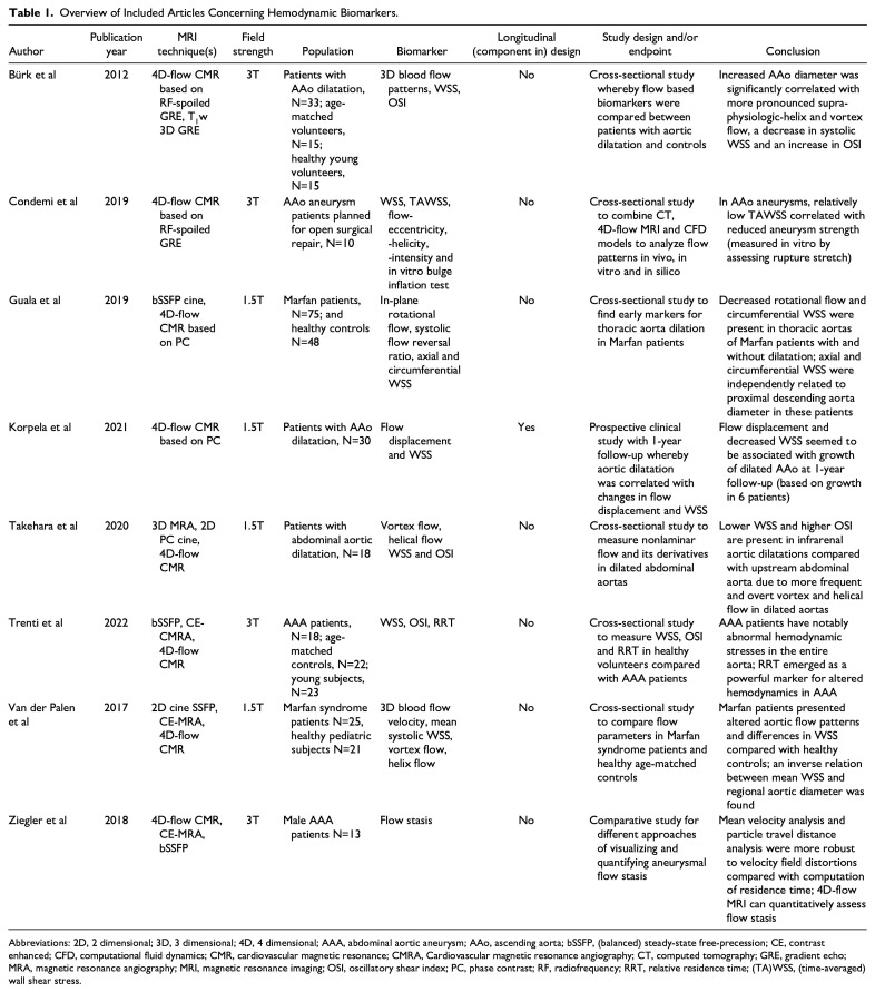
Results: A total of 1641 articles were identified, of which 21 were included in the scoping review. Quantitative MRI-derived biomarkers were categorized into hemodynamic (8 studies), wall (5 studies) and molecular biomarkers (8 studies). Fifteen studies included patients and/or healthy human subjects. Animal models were investigated in the other 6 studies. A cross-sectional study design was the most common, whereas 5 animal studies had a longitudinal component and 2 studies including patients had a prospective design. A promising hemodynamic biomarker is wall shear stress (WSS), which is estimated based on 4D-flow MRI. Molecular biomarkers enable the assessment of inflammatory and wall deterioration processes. The ADAMTS4-specific molecular magnetic resonance (MR) probe showed potential to predict abdominal aortic aneurysm (AAA) formation and rupture in a murine model. Wall biomarkers assessed using dynamic contrast-enhanced (DCE) MRI showed great potential for assessing AAA progression independent of the maximum diameter.
Conclusion: This scoping review provides an overview of quantitative MRI techniques studied and the biomarkers derived from them to assess aortic aneurysm progression and rupture risk. Longitudinal studies are needed to validate the causal relationships between the identified biomarkers and aneurysm growth, rupture, or repair. In the future, quantitative MRI could play an important role in the personalized risk assessment of aortic aneurysm rupture.Clinical ImpactThe currently used maximum aneurysm diameter fails to accurately assess the multifactorial pathology of an aortic aneurysm and precisely predicts rupture in a patient-specific manner. Quantitative magnetic resonance imaging (MRI) enables the detection of various quantitative parameters involved in aneurysm progression and subsequent rupture. This scoping review provides an overview of the studied quantitative MRI techniques, the biomarkers derived from them, and recommendations for future research needed for the implementation of these biomarkers. Ultimately, quantitative MRI could facilitate personalized risk assessment for patients with aortic aneurysms, thereby reducing untimely repairs and improving rupture prevention.
期刊介绍:
The Journal of Endovascular Therapy (formerly the Journal of Endovascular Surgery) was established in 1994 as a forum for all physicians, scientists, and allied healthcare professionals who are engaged or interested in peripheral endovascular techniques and technology. An official publication of the International Society of Endovascular Specialists (ISEVS), the Journal of Endovascular Therapy publishes peer-reviewed articles of interest to clinicians and researchers in the field of peripheral endovascular interventions.

 求助内容:
求助内容: 应助结果提醒方式:


