{"title":"TET2的异常表达是精索静脉曲张DNA甲基化不足的原因。","authors":"Hengameh Taghian Dinani, Nushin Naderi, Marziyeh Tavalaee, Farzaneh Rabiee, Mohammad Hossein Nasr-Esfahani","doi":"10.22074/cellj.2023.2000170.1284","DOIUrl":null,"url":null,"abstract":"<p><strong>Objective: </strong>Epigenetic modifications such as DNA methylation play a key role in male infertility etiology. This study aimed to explore the global DNA methylation status in testicular spermatogenic cells of varicocele-induced rats and consider their semen quality, with a focus on key epigenetic marks, namely 5-methylcytosine (5-mC) and 5-hydroxymethylcytosine (5-hmC), as well as the mRNA and proteins of ten-eleven translocation (TET) methylcytosine dioxygenases 1-3.</p><p><strong>Materials and methods: </strong>In this experimental study, 24 mature male Wistar rats (8 in each group) were assigned amongst the control, sham, and varicocele groups. Sperm quality was assessed, and DNA methylation patterns of testicular spermatogenic cells were investigated using reverse transcription-polymerase chain reaction (RT-PCR), western blot, and immunofluorescence techniques.</p><p><strong>Results: </strong>Sperm parameters, chromatin and DNA integrity were significantly lower, and sperm lipid peroxidation significantly increased in varicocele-induced rats in comparison with control rats. During spermatogenesis in rat testis, 5-mC and 5-hmC epigenetic marks, and TET1-3 mRNA and proteins were expressed. In contrast to the 5-mC fluorescent signal which was presented in all testicular cells, the 5-hmC fluorescent signal was presented exclusively in spermatogonia and a few spermatids. In varicocele-induced rats, the 5-mC signal decreased in all cells within the tubules, whereas a strong signal of 5-hmC was detected in seminiferous tubules compared to the control group. As well, the levels of TET2 mRNA and protein expression were significantly upregulated in varicocele-induced rats in comparison with the control group. Also, our results showed that the varicocele-induced animals exhibited strong fluorescent signals of TET1-3 in testicular cells, whereas weak fluorescent signals were identified in the seminiferous tubules of the control animals.</p><p><strong>Conclusion: </strong>Consequently, we showed TET2 upregulation and the 5-hmC gain at testicular levels are associated with varicocele and sperm quality decline, and therefore they can be exploited as potential biomarkers of spermatogenesis.</p>","PeriodicalId":49224,"journal":{"name":"Cell Journal","volume":"25 10","pages":"706-716"},"PeriodicalIF":1.7000,"publicationDate":"2023-10-09","publicationTypes":"Journal Article","fieldsOfStudy":null,"isOpenAccess":false,"openAccessPdf":"https://ftp.ncbi.nlm.nih.gov/pub/pmc/oa_pdf/13/9b/Cell-J-25-706.PMC10591265.pdf","citationCount":"0","resultStr":"{\"title\":\"Aberrant Expression of TET2 Accounts for DNA Hypomethylation in Varicocele.\",\"authors\":\"Hengameh Taghian Dinani, Nushin Naderi, Marziyeh Tavalaee, Farzaneh Rabiee, Mohammad Hossein Nasr-Esfahani\",\"doi\":\"10.22074/cellj.2023.2000170.1284\",\"DOIUrl\":null,\"url\":null,\"abstract\":\"<p><strong>Objective: </strong>Epigenetic modifications such as DNA methylation play a key role in male infertility etiology. This study aimed to explore the global DNA methylation status in testicular spermatogenic cells of varicocele-induced rats and consider their semen quality, with a focus on key epigenetic marks, namely 5-methylcytosine (5-mC) and 5-hydroxymethylcytosine (5-hmC), as well as the mRNA and proteins of ten-eleven translocation (TET) methylcytosine dioxygenases 1-3.</p><p><strong>Materials and methods: </strong>In this experimental study, 24 mature male Wistar rats (8 in each group) were assigned amongst the control, sham, and varicocele groups. Sperm quality was assessed, and DNA methylation patterns of testicular spermatogenic cells were investigated using reverse transcription-polymerase chain reaction (RT-PCR), western blot, and immunofluorescence techniques.</p><p><strong>Results: </strong>Sperm parameters, chromatin and DNA integrity were significantly lower, and sperm lipid peroxidation significantly increased in varicocele-induced rats in comparison with control rats. During spermatogenesis in rat testis, 5-mC and 5-hmC epigenetic marks, and TET1-3 mRNA and proteins were expressed. In contrast to the 5-mC fluorescent signal which was presented in all testicular cells, the 5-hmC fluorescent signal was presented exclusively in spermatogonia and a few spermatids. In varicocele-induced rats, the 5-mC signal decreased in all cells within the tubules, whereas a strong signal of 5-hmC was detected in seminiferous tubules compared to the control group. As well, the levels of TET2 mRNA and protein expression were significantly upregulated in varicocele-induced rats in comparison with the control group. Also, our results showed that the varicocele-induced animals exhibited strong fluorescent signals of TET1-3 in testicular cells, whereas weak fluorescent signals were identified in the seminiferous tubules of the control animals.</p><p><strong>Conclusion: </strong>Consequently, we showed TET2 upregulation and the 5-hmC gain at testicular levels are associated with varicocele and sperm quality decline, and therefore they can be exploited as potential biomarkers of spermatogenesis.</p>\",\"PeriodicalId\":49224,\"journal\":{\"name\":\"Cell Journal\",\"volume\":\"25 10\",\"pages\":\"706-716\"},\"PeriodicalIF\":1.7000,\"publicationDate\":\"2023-10-09\",\"publicationTypes\":\"Journal Article\",\"fieldsOfStudy\":null,\"isOpenAccess\":false,\"openAccessPdf\":\"https://ftp.ncbi.nlm.nih.gov/pub/pmc/oa_pdf/13/9b/Cell-J-25-706.PMC10591265.pdf\",\"citationCount\":\"0\",\"resultStr\":null,\"platform\":\"Semanticscholar\",\"paperid\":null,\"PeriodicalName\":\"Cell Journal\",\"FirstCategoryId\":\"99\",\"ListUrlMain\":\"https://doi.org/10.22074/cellj.2023.2000170.1284\",\"RegionNum\":4,\"RegionCategory\":\"生物学\",\"ArticlePicture\":[],\"TitleCN\":null,\"AbstractTextCN\":null,\"PMCID\":null,\"EPubDate\":\"\",\"PubModel\":\"\",\"JCR\":\"Q4\",\"JCRName\":\"CELL BIOLOGY\",\"Score\":null,\"Total\":0}","platform":"Semanticscholar","paperid":null,"PeriodicalName":"Cell Journal","FirstCategoryId":"99","ListUrlMain":"https://doi.org/10.22074/cellj.2023.2000170.1284","RegionNum":4,"RegionCategory":"生物学","ArticlePicture":[],"TitleCN":null,"AbstractTextCN":null,"PMCID":null,"EPubDate":"","PubModel":"","JCR":"Q4","JCRName":"CELL BIOLOGY","Score":null,"Total":0}
Aberrant Expression of TET2 Accounts for DNA Hypomethylation in Varicocele.
Objective: Epigenetic modifications such as DNA methylation play a key role in male infertility etiology. This study aimed to explore the global DNA methylation status in testicular spermatogenic cells of varicocele-induced rats and consider their semen quality, with a focus on key epigenetic marks, namely 5-methylcytosine (5-mC) and 5-hydroxymethylcytosine (5-hmC), as well as the mRNA and proteins of ten-eleven translocation (TET) methylcytosine dioxygenases 1-3.
Materials and methods: In this experimental study, 24 mature male Wistar rats (8 in each group) were assigned amongst the control, sham, and varicocele groups. Sperm quality was assessed, and DNA methylation patterns of testicular spermatogenic cells were investigated using reverse transcription-polymerase chain reaction (RT-PCR), western blot, and immunofluorescence techniques.
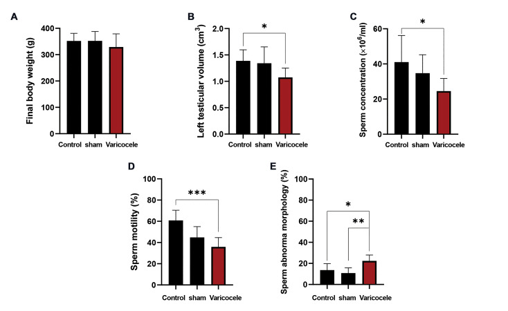
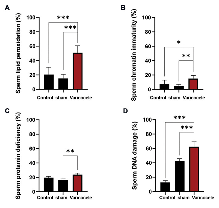
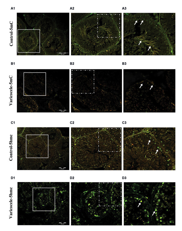
Results: Sperm parameters, chromatin and DNA integrity were significantly lower, and sperm lipid peroxidation significantly increased in varicocele-induced rats in comparison with control rats. During spermatogenesis in rat testis, 5-mC and 5-hmC epigenetic marks, and TET1-3 mRNA and proteins were expressed. In contrast to the 5-mC fluorescent signal which was presented in all testicular cells, the 5-hmC fluorescent signal was presented exclusively in spermatogonia and a few spermatids. In varicocele-induced rats, the 5-mC signal decreased in all cells within the tubules, whereas a strong signal of 5-hmC was detected in seminiferous tubules compared to the control group. As well, the levels of TET2 mRNA and protein expression were significantly upregulated in varicocele-induced rats in comparison with the control group. Also, our results showed that the varicocele-induced animals exhibited strong fluorescent signals of TET1-3 in testicular cells, whereas weak fluorescent signals were identified in the seminiferous tubules of the control animals.
Conclusion: Consequently, we showed TET2 upregulation and the 5-hmC gain at testicular levels are associated with varicocele and sperm quality decline, and therefore they can be exploited as potential biomarkers of spermatogenesis.
期刊介绍:
The “Cell Journal (Yakhteh)“, formerly published as “Yakhteh Medical Journal”, is a quarterly English publication of Royan Institute. This journal focuses on topics relevant to cellular and molecular scientific areas, besides other related fields. The Cell J has been certified by Ministry of Culture and Islamic Guidance in 1999 and was accredited as a scientific and research journal by HBI (Health and Biomedical Information) Journal Accreditation Commission in 2000 which is an open access journal.
 求助内容:
求助内容: 应助结果提醒方式:


