{"title":"铋铁氧体铁电响应的动态x射线衍射成像","authors":"Nouamane Laanait, Wittawat Saenrang, Hua Zhou, Chang-Beom Eom, Zhan Zhang","doi":"10.1186/s40679-017-0044-3","DOIUrl":null,"url":null,"abstract":"<p>X-ray diffraction imaging is rapidly emerging as a powerful technique by which one can capture the local structure of crystalline materials at the nano- and meso-scale. Here, we present investigations of the dynamic structure of epitaxial monodomain BiFeO<sub>3</sub> thin-films using a novel full-field Bragg diffraction imaging modality. By taking advantage of the depth penetration of hard X-rays and their exquisite sensitivity to the atomic structure, we imaged in situ and in operando, the electric field-driven structural responses of buried BiFeO<sub>3</sub> epitaxial thin-films in micro-capacitor devices, with sub-100?nm lateral resolution. These imaging investigations were carried out at acquisition frame rates that reached up to 20?Hz and data transfer rates of 40?MB/s, while accessing diffraction contrast that is sensitive to the entire three-dimensional unit cell configuration. We mined these large datasets for material responses by employing matrix decomposition techniques, such as independent component analysis. We found that this statistical approach allows the extraction of the salient physical properties of the ferroelectric response of the material, such as coercive fields and transient spatiotemporal modulations in their piezoelectric response, and also facilitates their decoupling from extrinsic sources that are instrument specific.</p>","PeriodicalId":460,"journal":{"name":"Advanced Structural and Chemical Imaging","volume":"3 1","pages":""},"PeriodicalIF":3.5600,"publicationDate":"2017-03-21","publicationTypes":"Journal Article","fieldsOfStudy":null,"isOpenAccess":false,"openAccessPdf":"https://sci-hub-pdf.com/10.1186/s40679-017-0044-3","citationCount":"9","resultStr":"{\"title\":\"Dynamic X-ray diffraction imaging of the ferroelectric response in bismuth ferrite\",\"authors\":\"Nouamane Laanait, Wittawat Saenrang, Hua Zhou, Chang-Beom Eom, Zhan Zhang\",\"doi\":\"10.1186/s40679-017-0044-3\",\"DOIUrl\":null,\"url\":null,\"abstract\":\"<p>X-ray diffraction imaging is rapidly emerging as a powerful technique by which one can capture the local structure of crystalline materials at the nano- and meso-scale. Here, we present investigations of the dynamic structure of epitaxial monodomain BiFeO<sub>3</sub> thin-films using a novel full-field Bragg diffraction imaging modality. By taking advantage of the depth penetration of hard X-rays and their exquisite sensitivity to the atomic structure, we imaged in situ and in operando, the electric field-driven structural responses of buried BiFeO<sub>3</sub> epitaxial thin-films in micro-capacitor devices, with sub-100?nm lateral resolution. These imaging investigations were carried out at acquisition frame rates that reached up to 20?Hz and data transfer rates of 40?MB/s, while accessing diffraction contrast that is sensitive to the entire three-dimensional unit cell configuration. We mined these large datasets for material responses by employing matrix decomposition techniques, such as independent component analysis. We found that this statistical approach allows the extraction of the salient physical properties of the ferroelectric response of the material, such as coercive fields and transient spatiotemporal modulations in their piezoelectric response, and also facilitates their decoupling from extrinsic sources that are instrument specific.</p>\",\"PeriodicalId\":460,\"journal\":{\"name\":\"Advanced Structural and Chemical Imaging\",\"volume\":\"3 1\",\"pages\":\"\"},\"PeriodicalIF\":3.5600,\"publicationDate\":\"2017-03-21\",\"publicationTypes\":\"Journal Article\",\"fieldsOfStudy\":null,\"isOpenAccess\":false,\"openAccessPdf\":\"https://sci-hub-pdf.com/10.1186/s40679-017-0044-3\",\"citationCount\":\"9\",\"resultStr\":null,\"platform\":\"Semanticscholar\",\"paperid\":null,\"PeriodicalName\":\"Advanced Structural and Chemical Imaging\",\"FirstCategoryId\":\"1085\",\"ListUrlMain\":\"https://link.springer.com/article/10.1186/s40679-017-0044-3\",\"RegionNum\":0,\"RegionCategory\":null,\"ArticlePicture\":[],\"TitleCN\":null,\"AbstractTextCN\":null,\"PMCID\":null,\"EPubDate\":\"\",\"PubModel\":\"\",\"JCR\":\"Q1\",\"JCRName\":\"Medicine\",\"Score\":null,\"Total\":0}","platform":"Semanticscholar","paperid":null,"PeriodicalName":"Advanced Structural and Chemical Imaging","FirstCategoryId":"1085","ListUrlMain":"https://link.springer.com/article/10.1186/s40679-017-0044-3","RegionNum":0,"RegionCategory":null,"ArticlePicture":[],"TitleCN":null,"AbstractTextCN":null,"PMCID":null,"EPubDate":"","PubModel":"","JCR":"Q1","JCRName":"Medicine","Score":null,"Total":0}
Dynamic X-ray diffraction imaging of the ferroelectric response in bismuth ferrite
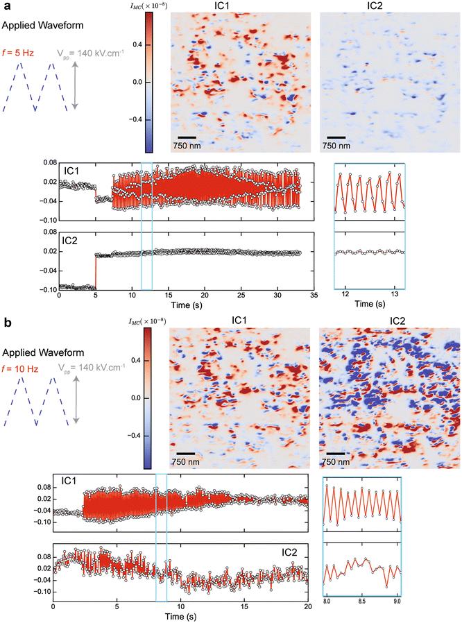
X-ray diffraction imaging is rapidly emerging as a powerful technique by which one can capture the local structure of crystalline materials at the nano- and meso-scale. Here, we present investigations of the dynamic structure of epitaxial monodomain BiFeO3 thin-films using a novel full-field Bragg diffraction imaging modality. By taking advantage of the depth penetration of hard X-rays and their exquisite sensitivity to the atomic structure, we imaged in situ and in operando, the electric field-driven structural responses of buried BiFeO3 epitaxial thin-films in micro-capacitor devices, with sub-100?nm lateral resolution. These imaging investigations were carried out at acquisition frame rates that reached up to 20?Hz and data transfer rates of 40?MB/s, while accessing diffraction contrast that is sensitive to the entire three-dimensional unit cell configuration. We mined these large datasets for material responses by employing matrix decomposition techniques, such as independent component analysis. We found that this statistical approach allows the extraction of the salient physical properties of the ferroelectric response of the material, such as coercive fields and transient spatiotemporal modulations in their piezoelectric response, and also facilitates their decoupling from extrinsic sources that are instrument specific.

 求助内容:
求助内容: 应助结果提醒方式:


