Carol B. Johnson, Zheng Long, Zhiping Luo, Rahamthulla S. Shaik, Min Woo Sung, Stanislav Vitha, Andreas Holzenburg
{"title":"拟南芥叶绿体中FtsZ微环的原位结构","authors":"Carol B. Johnson, Zheng Long, Zhiping Luo, Rahamthulla S. Shaik, Min Woo Sung, Stanislav Vitha, Andreas Holzenburg","doi":"10.1186/s40679-015-0013-7","DOIUrl":null,"url":null,"abstract":"<p>Chloroplasts are essential plant organelles that divide by binary fission through a coordinated ring-shaped division machinery located both on the outside and inside of the chloroplast. The first step in chloroplast division is the assembly of an internal division ring (Z-ring) that is composed of the key filamentous chloroplast division proteins FtsZ1 and FtsZ2. How the individual FtsZ filaments assemble into higher-order structures to form the dividing Z-ring is not well understood and the most detailed insights have so far been gleaned from prokaryotic FtsZ. Here, we present in situ data of chloroplast FtsZ making use of a smaller ring-like FtsZ assembly termed mini-rings that form under well-defined conditions. Structured illumination microscopy (SIM) permitted their mean diameter to be determined as 208?nm and also showed that 68?% of these rings are terminally attached to linear FtsZ filaments. A correlative microscopy-compatible specimen preparation based on freeze substitution after high-pressure freezing is presented addressing the challenges such as autofluorescence and specific fluorescence attenuation. Transmission electron microscopy (TEM) and scanning TEM (STEM) imaging of thin sections exhibited ring-like densities that matched in size with the SIM data, and TEM tomography revealed insights into the molecular architecture of mini-rings demonstrating the following key features: (1) overall, a roughly bipartite split into a more ordered/curved and less ordered/curved half is readily discernible; (2) the density distribution in individual strands matches with the X-ray data, suggesting they constitute FtsZ protofilaments; (3) in the less ordered half of the ring, the protofilaments are able to assemble into higher-order structures such as double helices and supercoiled structures. Taken together, the data suggest that the state of existence of mini-rings could be described as metastable and their possible involvement in filament storage and Z-ring assembly is discussed.</p>","PeriodicalId":460,"journal":{"name":"Advanced Structural and Chemical Imaging","volume":"1 1","pages":""},"PeriodicalIF":3.5600,"publicationDate":"2015-09-17","publicationTypes":"Journal Article","fieldsOfStudy":null,"isOpenAccess":false,"openAccessPdf":"https://sci-hub-pdf.com/10.1186/s40679-015-0013-7","citationCount":"4","resultStr":"{\"title\":\"In situ structure of FtsZ mini-rings in Arabidopsis chloroplasts\",\"authors\":\"Carol B. Johnson, Zheng Long, Zhiping Luo, Rahamthulla S. Shaik, Min Woo Sung, Stanislav Vitha, Andreas Holzenburg\",\"doi\":\"10.1186/s40679-015-0013-7\",\"DOIUrl\":null,\"url\":null,\"abstract\":\"<p>Chloroplasts are essential plant organelles that divide by binary fission through a coordinated ring-shaped division machinery located both on the outside and inside of the chloroplast. The first step in chloroplast division is the assembly of an internal division ring (Z-ring) that is composed of the key filamentous chloroplast division proteins FtsZ1 and FtsZ2. How the individual FtsZ filaments assemble into higher-order structures to form the dividing Z-ring is not well understood and the most detailed insights have so far been gleaned from prokaryotic FtsZ. Here, we present in situ data of chloroplast FtsZ making use of a smaller ring-like FtsZ assembly termed mini-rings that form under well-defined conditions. Structured illumination microscopy (SIM) permitted their mean diameter to be determined as 208?nm and also showed that 68?% of these rings are terminally attached to linear FtsZ filaments. A correlative microscopy-compatible specimen preparation based on freeze substitution after high-pressure freezing is presented addressing the challenges such as autofluorescence and specific fluorescence attenuation. Transmission electron microscopy (TEM) and scanning TEM (STEM) imaging of thin sections exhibited ring-like densities that matched in size with the SIM data, and TEM tomography revealed insights into the molecular architecture of mini-rings demonstrating the following key features: (1) overall, a roughly bipartite split into a more ordered/curved and less ordered/curved half is readily discernible; (2) the density distribution in individual strands matches with the X-ray data, suggesting they constitute FtsZ protofilaments; (3) in the less ordered half of the ring, the protofilaments are able to assemble into higher-order structures such as double helices and supercoiled structures. Taken together, the data suggest that the state of existence of mini-rings could be described as metastable and their possible involvement in filament storage and Z-ring assembly is discussed.</p>\",\"PeriodicalId\":460,\"journal\":{\"name\":\"Advanced Structural and Chemical Imaging\",\"volume\":\"1 1\",\"pages\":\"\"},\"PeriodicalIF\":3.5600,\"publicationDate\":\"2015-09-17\",\"publicationTypes\":\"Journal Article\",\"fieldsOfStudy\":null,\"isOpenAccess\":false,\"openAccessPdf\":\"https://sci-hub-pdf.com/10.1186/s40679-015-0013-7\",\"citationCount\":\"4\",\"resultStr\":null,\"platform\":\"Semanticscholar\",\"paperid\":null,\"PeriodicalName\":\"Advanced Structural and Chemical Imaging\",\"FirstCategoryId\":\"1085\",\"ListUrlMain\":\"https://link.springer.com/article/10.1186/s40679-015-0013-7\",\"RegionNum\":0,\"RegionCategory\":null,\"ArticlePicture\":[],\"TitleCN\":null,\"AbstractTextCN\":null,\"PMCID\":null,\"EPubDate\":\"\",\"PubModel\":\"\",\"JCR\":\"Q1\",\"JCRName\":\"Medicine\",\"Score\":null,\"Total\":0}","platform":"Semanticscholar","paperid":null,"PeriodicalName":"Advanced Structural and Chemical Imaging","FirstCategoryId":"1085","ListUrlMain":"https://link.springer.com/article/10.1186/s40679-015-0013-7","RegionNum":0,"RegionCategory":null,"ArticlePicture":[],"TitleCN":null,"AbstractTextCN":null,"PMCID":null,"EPubDate":"","PubModel":"","JCR":"Q1","JCRName":"Medicine","Score":null,"Total":0}
In situ structure of FtsZ mini-rings in Arabidopsis chloroplasts
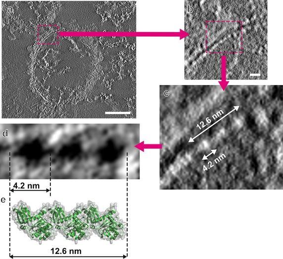
Chloroplasts are essential plant organelles that divide by binary fission through a coordinated ring-shaped division machinery located both on the outside and inside of the chloroplast. The first step in chloroplast division is the assembly of an internal division ring (Z-ring) that is composed of the key filamentous chloroplast division proteins FtsZ1 and FtsZ2. How the individual FtsZ filaments assemble into higher-order structures to form the dividing Z-ring is not well understood and the most detailed insights have so far been gleaned from prokaryotic FtsZ. Here, we present in situ data of chloroplast FtsZ making use of a smaller ring-like FtsZ assembly termed mini-rings that form under well-defined conditions. Structured illumination microscopy (SIM) permitted their mean diameter to be determined as 208?nm and also showed that 68?% of these rings are terminally attached to linear FtsZ filaments. A correlative microscopy-compatible specimen preparation based on freeze substitution after high-pressure freezing is presented addressing the challenges such as autofluorescence and specific fluorescence attenuation. Transmission electron microscopy (TEM) and scanning TEM (STEM) imaging of thin sections exhibited ring-like densities that matched in size with the SIM data, and TEM tomography revealed insights into the molecular architecture of mini-rings demonstrating the following key features: (1) overall, a roughly bipartite split into a more ordered/curved and less ordered/curved half is readily discernible; (2) the density distribution in individual strands matches with the X-ray data, suggesting they constitute FtsZ protofilaments; (3) in the less ordered half of the ring, the protofilaments are able to assemble into higher-order structures such as double helices and supercoiled structures. Taken together, the data suggest that the state of existence of mini-rings could be described as metastable and their possible involvement in filament storage and Z-ring assembly is discussed.

 求助内容:
求助内容: 应助结果提醒方式:


