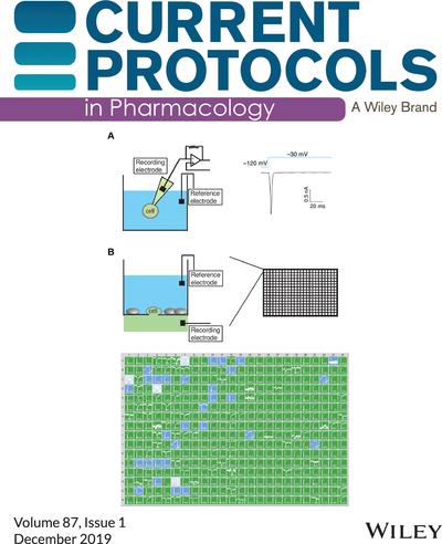
{"title":"发布信息TOC","authors":"","doi":"10.1002/cpph.53","DOIUrl":null,"url":null,"abstract":"<p><b>Cover</b>: In Liu et al. (https://doi.org/10.1002/cpph.69), the image shows a comparison between conventional manual patch clamp and automated electrophysiology. (<b>A</b>) Schematic of manual patch clamp recording (left panel) under whole-cell configuration. A single cell is recorded using a glass recording pipette. Compounds can be applied to the extracellular solution (blue), whereas the artificial intracellular solution (green) is contained in the pipette. Right panel: voltage pulse protocol and representative transient inward Nav1.7 channel current. (<b>B</b>) Schematic of planar array–based automated electrophysiology experiment. A single cell is recorded through a micropore in a single well (upper left) from a chip containing 384 wells (upper right). Both extracellular (blue) and intracellular (green) solutions can be changed by perfusion. Lower panel: representative result from one Syncropatch run. Wells were color coded based on seal resistance. Resistance, cells: green, >0.5 GΩ, 350/384; blue, 100 to 500 MΩ, 30/384; white, <100 MΩ, 4/384. Current traces within each well were autoscaled for maximum display.\n\n <figure>\n <div><picture>\n <source></source></picture><p></p>\n </div>\n </figure></p>","PeriodicalId":10871,"journal":{"name":"Current Protocols in Pharmacology","volume":"87 1","pages":""},"PeriodicalIF":0.0000,"publicationDate":"2019-12-22","publicationTypes":"Journal Article","fieldsOfStudy":null,"isOpenAccess":false,"openAccessPdf":"https://sci-hub-pdf.com/10.1002/cpph.53","citationCount":"0","resultStr":"{\"title\":\"Issue Information TOC\",\"authors\":\"\",\"doi\":\"10.1002/cpph.53\",\"DOIUrl\":null,\"url\":null,\"abstract\":\"<p><b>Cover</b>: In Liu et al. (https://doi.org/10.1002/cpph.69), the image shows a comparison between conventional manual patch clamp and automated electrophysiology. (<b>A</b>) Schematic of manual patch clamp recording (left panel) under whole-cell configuration. A single cell is recorded using a glass recording pipette. Compounds can be applied to the extracellular solution (blue), whereas the artificial intracellular solution (green) is contained in the pipette. Right panel: voltage pulse protocol and representative transient inward Nav1.7 channel current. (<b>B</b>) Schematic of planar array–based automated electrophysiology experiment. A single cell is recorded through a micropore in a single well (upper left) from a chip containing 384 wells (upper right). Both extracellular (blue) and intracellular (green) solutions can be changed by perfusion. Lower panel: representative result from one Syncropatch run. Wells were color coded based on seal resistance. Resistance, cells: green, >0.5 GΩ, 350/384; blue, 100 to 500 MΩ, 30/384; white, <100 MΩ, 4/384. Current traces within each well were autoscaled for maximum display.\\n\\n <figure>\\n <div><picture>\\n <source></source></picture><p></p>\\n </div>\\n </figure></p>\",\"PeriodicalId\":10871,\"journal\":{\"name\":\"Current Protocols in Pharmacology\",\"volume\":\"87 1\",\"pages\":\"\"},\"PeriodicalIF\":0.0000,\"publicationDate\":\"2019-12-22\",\"publicationTypes\":\"Journal Article\",\"fieldsOfStudy\":null,\"isOpenAccess\":false,\"openAccessPdf\":\"https://sci-hub-pdf.com/10.1002/cpph.53\",\"citationCount\":\"0\",\"resultStr\":null,\"platform\":\"Semanticscholar\",\"paperid\":null,\"PeriodicalName\":\"Current Protocols in Pharmacology\",\"FirstCategoryId\":\"1085\",\"ListUrlMain\":\"https://onlinelibrary.wiley.com/doi/10.1002/cpph.53\",\"RegionNum\":0,\"RegionCategory\":null,\"ArticlePicture\":[],\"TitleCN\":null,\"AbstractTextCN\":null,\"PMCID\":null,\"EPubDate\":\"\",\"PubModel\":\"\",\"JCR\":\"Q2\",\"JCRName\":\"Pharmacology, Toxicology and Pharmaceutics\",\"Score\":null,\"Total\":0}","platform":"Semanticscholar","paperid":null,"PeriodicalName":"Current Protocols in Pharmacology","FirstCategoryId":"1085","ListUrlMain":"https://onlinelibrary.wiley.com/doi/10.1002/cpph.53","RegionNum":0,"RegionCategory":null,"ArticlePicture":[],"TitleCN":null,"AbstractTextCN":null,"PMCID":null,"EPubDate":"","PubModel":"","JCR":"Q2","JCRName":"Pharmacology, Toxicology and Pharmaceutics","Score":null,"Total":0}
Cover: In Liu et al. (https://doi.org/10.1002/cpph.69), the image shows a comparison between conventional manual patch clamp and automated electrophysiology. (A) Schematic of manual patch clamp recording (left panel) under whole-cell configuration. A single cell is recorded using a glass recording pipette. Compounds can be applied to the extracellular solution (blue), whereas the artificial intracellular solution (green) is contained in the pipette. Right panel: voltage pulse protocol and representative transient inward Nav1.7 channel current. (B) Schematic of planar array–based automated electrophysiology experiment. A single cell is recorded through a micropore in a single well (upper left) from a chip containing 384 wells (upper right). Both extracellular (blue) and intracellular (green) solutions can be changed by perfusion. Lower panel: representative result from one Syncropatch run. Wells were color coded based on seal resistance. Resistance, cells: green, >0.5 GΩ, 350/384; blue, 100 to 500 MΩ, 30/384; white, <100 MΩ, 4/384. Current traces within each well were autoscaled for maximum display.

 求助内容:
求助内容: 应助结果提醒方式:


