Joseph D Buehler, Cylaina E Bird, Milan R Savani, Lauren C Gattie, William H Hicks, Michael M Levitt, Mohamad El Shami, Kimmo J Hatanpaa, Timothy E Richardson, Samuel K McBrayer, Kalil G Abdullah
{"title":"用快速活细胞显微镜对癌症类器官生存能力的半自动计算评估","authors":"Joseph D Buehler, Cylaina E Bird, Milan R Savani, Lauren C Gattie, William H Hicks, Michael M Levitt, Mohamad El Shami, Kimmo J Hatanpaa, Timothy E Richardson, Samuel K McBrayer, Kalil G Abdullah","doi":"10.1177/11769351221100754","DOIUrl":null,"url":null,"abstract":"<p><p>The creation of patient-derived cancer organoids represents a key advance in preclinical modeling and has recently been applied to a variety of human solid tumor types. However, conventional methods used to assess in vivo tumor tissue treatment response are poorly suited for the evaluation of cancer organoids because they are time-intensive and involve tissue destruction. To address this issue, we established a suite of 3-dimensional patient-derived glioma organoids, treated them with chemoradiotherapy, stained organoids with non-toxic cell dyes, and imaged them using a rapid laser scanning confocal microscopy method termed \"Apex Imaging.\" We then developed and tested a fragmentation algorithm to quantify heterogeneity in the topography of the organoids as a potential surrogate marker of viability. This algorithm, SSDquant, provides a 3-dimensional visual representation of the organoid surface and a numerical measurement of the sum-squared distance (SSD) from the derived mass center of the organoid. We tested whether SSD scores correlate with traditional immunohistochemistry-derived cell viability markers (cellularity and cleaved caspase 3 expression) and observed statistically significant associations between them using linear regression analysis. Our work describes a quantitative, non-invasive approach for the serial measurement of patient-derived cancer organoid viability, thus opening new avenues for the application of these models to studies of cancer biology and therapy.</p>","PeriodicalId":35418,"journal":{"name":"Cancer Informatics","volume":"21 1","pages":"11769351221100754"},"PeriodicalIF":2.5000,"publicationDate":"2022-05-26","publicationTypes":"Journal Article","fieldsOfStudy":null,"isOpenAccess":false,"openAccessPdf":"https://www.ncbi.nlm.nih.gov/pmc/articles/PMC9150230/pdf/","citationCount":"0","resultStr":"{\"title\":\"Semi-Automated Computational Assessment of Cancer Organoid Viability Using Rapid Live-Cell Microscopy.\",\"authors\":\"Joseph D Buehler, Cylaina E Bird, Milan R Savani, Lauren C Gattie, William H Hicks, Michael M Levitt, Mohamad El Shami, Kimmo J Hatanpaa, Timothy E Richardson, Samuel K McBrayer, Kalil G Abdullah\",\"doi\":\"10.1177/11769351221100754\",\"DOIUrl\":null,\"url\":null,\"abstract\":\"<p><p>The creation of patient-derived cancer organoids represents a key advance in preclinical modeling and has recently been applied to a variety of human solid tumor types. However, conventional methods used to assess in vivo tumor tissue treatment response are poorly suited for the evaluation of cancer organoids because they are time-intensive and involve tissue destruction. To address this issue, we established a suite of 3-dimensional patient-derived glioma organoids, treated them with chemoradiotherapy, stained organoids with non-toxic cell dyes, and imaged them using a rapid laser scanning confocal microscopy method termed \\\"Apex Imaging.\\\" We then developed and tested a fragmentation algorithm to quantify heterogeneity in the topography of the organoids as a potential surrogate marker of viability. This algorithm, SSDquant, provides a 3-dimensional visual representation of the organoid surface and a numerical measurement of the sum-squared distance (SSD) from the derived mass center of the organoid. We tested whether SSD scores correlate with traditional immunohistochemistry-derived cell viability markers (cellularity and cleaved caspase 3 expression) and observed statistically significant associations between them using linear regression analysis. Our work describes a quantitative, non-invasive approach for the serial measurement of patient-derived cancer organoid viability, thus opening new avenues for the application of these models to studies of cancer biology and therapy.</p>\",\"PeriodicalId\":35418,\"journal\":{\"name\":\"Cancer Informatics\",\"volume\":\"21 1\",\"pages\":\"11769351221100754\"},\"PeriodicalIF\":2.5000,\"publicationDate\":\"2022-05-26\",\"publicationTypes\":\"Journal Article\",\"fieldsOfStudy\":null,\"isOpenAccess\":false,\"openAccessPdf\":\"https://www.ncbi.nlm.nih.gov/pmc/articles/PMC9150230/pdf/\",\"citationCount\":\"0\",\"resultStr\":null,\"platform\":\"Semanticscholar\",\"paperid\":null,\"PeriodicalName\":\"Cancer Informatics\",\"FirstCategoryId\":\"1085\",\"ListUrlMain\":\"https://doi.org/10.1177/11769351221100754\",\"RegionNum\":0,\"RegionCategory\":null,\"ArticlePicture\":[],\"TitleCN\":null,\"AbstractTextCN\":null,\"PMCID\":null,\"EPubDate\":\"2022/1/1 0:00:00\",\"PubModel\":\"eCollection\",\"JCR\":\"Q2\",\"JCRName\":\"MATHEMATICAL & COMPUTATIONAL BIOLOGY\",\"Score\":null,\"Total\":0}","platform":"Semanticscholar","paperid":null,"PeriodicalName":"Cancer Informatics","FirstCategoryId":"1085","ListUrlMain":"https://doi.org/10.1177/11769351221100754","RegionNum":0,"RegionCategory":null,"ArticlePicture":[],"TitleCN":null,"AbstractTextCN":null,"PMCID":null,"EPubDate":"2022/1/1 0:00:00","PubModel":"eCollection","JCR":"Q2","JCRName":"MATHEMATICAL & COMPUTATIONAL BIOLOGY","Score":null,"Total":0}
Semi-Automated Computational Assessment of Cancer Organoid Viability Using Rapid Live-Cell Microscopy.
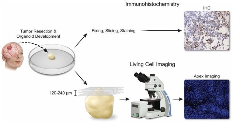
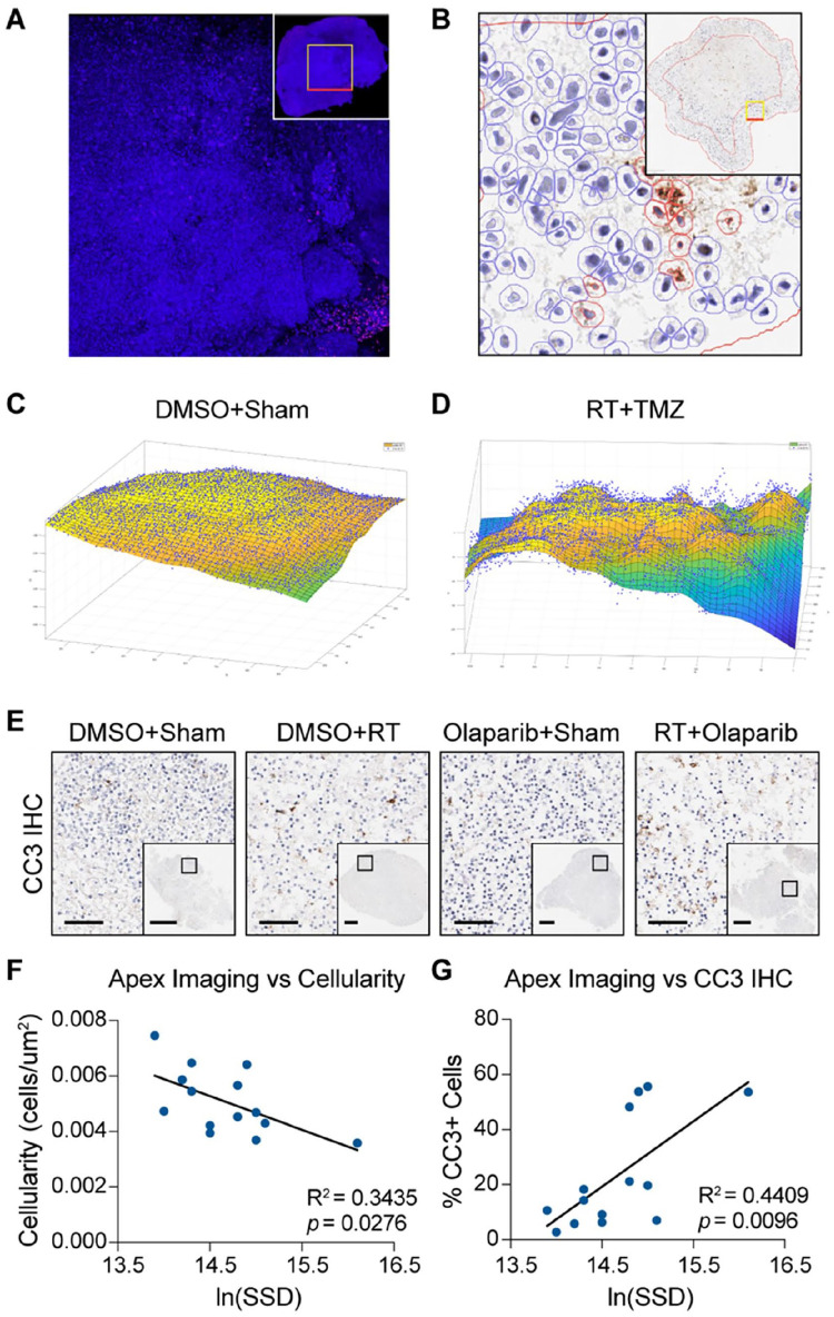
The creation of patient-derived cancer organoids represents a key advance in preclinical modeling and has recently been applied to a variety of human solid tumor types. However, conventional methods used to assess in vivo tumor tissue treatment response are poorly suited for the evaluation of cancer organoids because they are time-intensive and involve tissue destruction. To address this issue, we established a suite of 3-dimensional patient-derived glioma organoids, treated them with chemoradiotherapy, stained organoids with non-toxic cell dyes, and imaged them using a rapid laser scanning confocal microscopy method termed "Apex Imaging." We then developed and tested a fragmentation algorithm to quantify heterogeneity in the topography of the organoids as a potential surrogate marker of viability. This algorithm, SSDquant, provides a 3-dimensional visual representation of the organoid surface and a numerical measurement of the sum-squared distance (SSD) from the derived mass center of the organoid. We tested whether SSD scores correlate with traditional immunohistochemistry-derived cell viability markers (cellularity and cleaved caspase 3 expression) and observed statistically significant associations between them using linear regression analysis. Our work describes a quantitative, non-invasive approach for the serial measurement of patient-derived cancer organoid viability, thus opening new avenues for the application of these models to studies of cancer biology and therapy.
期刊介绍:
The field of cancer research relies on advances in many other disciplines, including omics technology, mass spectrometry, radio imaging, computer science, and biostatistics. Cancer Informatics provides open access to peer-reviewed high-quality manuscripts reporting bioinformatics analysis of molecular genetics and/or clinical data pertaining to cancer, emphasizing the use of machine learning, artificial intelligence, statistical algorithms, advanced imaging techniques, data visualization, and high-throughput technologies. As the leading journal dedicated exclusively to the report of the use of computational methods in cancer research and practice, Cancer Informatics leverages methodological improvements in systems biology, genomics, proteomics, metabolomics, and molecular biochemistry into the fields of cancer detection, treatment, classification, risk-prediction, prevention, outcome, and modeling.

 求助内容:
求助内容: 应助结果提醒方式:


