Yuan Zhuang, W. Binbin, Zhang Shaodong, Jun‐hua Li, H. Wan, Jie Feng, De-zhi Li, Song Liu
{"title":"面神经保留对神经再生及轮匝肌功能重建影响的实验研究","authors":"Yuan Zhuang, W. Binbin, Zhang Shaodong, Jun‐hua Li, H. Wan, Jie Feng, De-zhi Li, Song Liu","doi":"10.3760/CMA.J.ISSN.1001-2346.2020.02.017","DOIUrl":null,"url":null,"abstract":"Objective To investigate the effect of the regenerated facial nerve (FN) with varying degrees on the reconstruction of orbicularis function post facial-hypoglossal side-to-side neurorrhaphy (FN-HN) in the incomplete facial palsy model. Methods A total of 40 male adult Sprague Dawley rats were divided into 4 groups randomly. All rats were performed with crushed FN injury and FN-HN anastomosis. Three months later, the 4 groups were performed with FN cross section with different ratios at the proximal end to the injured site: no intervention in group A, 1/3 of cross section in group B, 2/3 of cross section in group C and transection in group D. The degree of closure of the affected-side eyelids in rats was assessed using the eye blink score, the muscle active potentials (MAPs) were used to detect the action potential and the area under the curve (AUC) of the rat′s orbicularis oculi muscle on the affected side, the number of positive neurons in the FN and HN nuclei were revealed by retrograde labeling, and the myelinated axons were counted on semi-thin sections of facial nerves and nerve grafts. Results For the eye blink scores, the 4 groups showed significant difference during the three months (F=12.47, 11.00, 10.61, 19.13, all P<0.05) and after the cross section (F=29.06, P<0.05). Moreover, the score in group D was lower than that in group A and B(both P<0.01). One week after the cross-section of FN, all 4 groups showed significant difference in amplitude and AUC of MAPs when we stimulated the proximal end of anastomosis site of facial nerve and the middle of the nerve graft (F=27.56, 11.86, 6.33, 4.65, all P<0.05). In addition, the amplitude and AUC of MAPs in group D were significantly lower than those in group A (both P<0.05). Among the 4 groups, retrograde labeling indicated significant difference in the number of positive neurons in the FN nucleus (F=6.52, P<0.05), and the neurons in group D were less than those in group A and B (both P<0.05). The counts of myelinated axons in both FN trunk and nerve graft in the 4 groups were significantly different (F=8.33, 6.35, both P<0.05). In particular, group A had less myelinated axons compared with group C and group D (both P<0.05). Conclusion Preserving structural integrity of facial nerve during FN-HN seems to have positive significance. Retention of at least 2/3 regenerated facial nerve is beneficial for the recovery of the orbicularis oculi muscle. Key words: Anastomosis, surgical; Facial nerve; Hypoglossal nerve; Orbicularis; Rats","PeriodicalId":10100,"journal":{"name":"中华神经外科杂志","volume":"36 1","pages":"188-193"},"PeriodicalIF":0.0000,"publicationDate":"2020-02-28","publicationTypes":"Journal Article","fieldsOfStudy":null,"isOpenAccess":false,"openAccessPdf":"","citationCount":"0","resultStr":"{\"title\":\"Experimental study on the effect of facial nerve retention on nerve regeneration and reconstruction of orbicularis muscle function\",\"authors\":\"Yuan Zhuang, W. Binbin, Zhang Shaodong, Jun‐hua Li, H. Wan, Jie Feng, De-zhi Li, Song Liu\",\"doi\":\"10.3760/CMA.J.ISSN.1001-2346.2020.02.017\",\"DOIUrl\":null,\"url\":null,\"abstract\":\"Objective To investigate the effect of the regenerated facial nerve (FN) with varying degrees on the reconstruction of orbicularis function post facial-hypoglossal side-to-side neurorrhaphy (FN-HN) in the incomplete facial palsy model. Methods A total of 40 male adult Sprague Dawley rats were divided into 4 groups randomly. All rats were performed with crushed FN injury and FN-HN anastomosis. Three months later, the 4 groups were performed with FN cross section with different ratios at the proximal end to the injured site: no intervention in group A, 1/3 of cross section in group B, 2/3 of cross section in group C and transection in group D. The degree of closure of the affected-side eyelids in rats was assessed using the eye blink score, the muscle active potentials (MAPs) were used to detect the action potential and the area under the curve (AUC) of the rat′s orbicularis oculi muscle on the affected side, the number of positive neurons in the FN and HN nuclei were revealed by retrograde labeling, and the myelinated axons were counted on semi-thin sections of facial nerves and nerve grafts. Results For the eye blink scores, the 4 groups showed significant difference during the three months (F=12.47, 11.00, 10.61, 19.13, all P<0.05) and after the cross section (F=29.06, P<0.05). Moreover, the score in group D was lower than that in group A and B(both P<0.01). One week after the cross-section of FN, all 4 groups showed significant difference in amplitude and AUC of MAPs when we stimulated the proximal end of anastomosis site of facial nerve and the middle of the nerve graft (F=27.56, 11.86, 6.33, 4.65, all P<0.05). In addition, the amplitude and AUC of MAPs in group D were significantly lower than those in group A (both P<0.05). Among the 4 groups, retrograde labeling indicated significant difference in the number of positive neurons in the FN nucleus (F=6.52, P<0.05), and the neurons in group D were less than those in group A and B (both P<0.05). The counts of myelinated axons in both FN trunk and nerve graft in the 4 groups were significantly different (F=8.33, 6.35, both P<0.05). In particular, group A had less myelinated axons compared with group C and group D (both P<0.05). Conclusion Preserving structural integrity of facial nerve during FN-HN seems to have positive significance. Retention of at least 2/3 regenerated facial nerve is beneficial for the recovery of the orbicularis oculi muscle. Key words: Anastomosis, surgical; Facial nerve; Hypoglossal nerve; Orbicularis; Rats\",\"PeriodicalId\":10100,\"journal\":{\"name\":\"中华神经外科杂志\",\"volume\":\"36 1\",\"pages\":\"188-193\"},\"PeriodicalIF\":0.0000,\"publicationDate\":\"2020-02-28\",\"publicationTypes\":\"Journal Article\",\"fieldsOfStudy\":null,\"isOpenAccess\":false,\"openAccessPdf\":\"\",\"citationCount\":\"0\",\"resultStr\":null,\"platform\":\"Semanticscholar\",\"paperid\":null,\"PeriodicalName\":\"中华神经外科杂志\",\"FirstCategoryId\":\"3\",\"ListUrlMain\":\"https://doi.org/10.3760/CMA.J.ISSN.1001-2346.2020.02.017\",\"RegionNum\":0,\"RegionCategory\":null,\"ArticlePicture\":[],\"TitleCN\":null,\"AbstractTextCN\":null,\"PMCID\":null,\"EPubDate\":\"\",\"PubModel\":\"\",\"JCR\":\"Q4\",\"JCRName\":\"Medicine\",\"Score\":null,\"Total\":0}","platform":"Semanticscholar","paperid":null,"PeriodicalName":"中华神经外科杂志","FirstCategoryId":"3","ListUrlMain":"https://doi.org/10.3760/CMA.J.ISSN.1001-2346.2020.02.017","RegionNum":0,"RegionCategory":null,"ArticlePicture":[],"TitleCN":null,"AbstractTextCN":null,"PMCID":null,"EPubDate":"","PubModel":"","JCR":"Q4","JCRName":"Medicine","Score":null,"Total":0}
Experimental study on the effect of facial nerve retention on nerve regeneration and reconstruction of orbicularis muscle function
Objective To investigate the effect of the regenerated facial nerve (FN) with varying degrees on the reconstruction of orbicularis function post facial-hypoglossal side-to-side neurorrhaphy (FN-HN) in the incomplete facial palsy model. Methods A total of 40 male adult Sprague Dawley rats were divided into 4 groups randomly. All rats were performed with crushed FN injury and FN-HN anastomosis. Three months later, the 4 groups were performed with FN cross section with different ratios at the proximal end to the injured site: no intervention in group A, 1/3 of cross section in group B, 2/3 of cross section in group C and transection in group D. The degree of closure of the affected-side eyelids in rats was assessed using the eye blink score, the muscle active potentials (MAPs) were used to detect the action potential and the area under the curve (AUC) of the rat′s orbicularis oculi muscle on the affected side, the number of positive neurons in the FN and HN nuclei were revealed by retrograde labeling, and the myelinated axons were counted on semi-thin sections of facial nerves and nerve grafts. Results For the eye blink scores, the 4 groups showed significant difference during the three months (F=12.47, 11.00, 10.61, 19.13, all P<0.05) and after the cross section (F=29.06, P<0.05). Moreover, the score in group D was lower than that in group A and B(both P<0.01). One week after the cross-section of FN, all 4 groups showed significant difference in amplitude and AUC of MAPs when we stimulated the proximal end of anastomosis site of facial nerve and the middle of the nerve graft (F=27.56, 11.86, 6.33, 4.65, all P<0.05). In addition, the amplitude and AUC of MAPs in group D were significantly lower than those in group A (both P<0.05). Among the 4 groups, retrograde labeling indicated significant difference in the number of positive neurons in the FN nucleus (F=6.52, P<0.05), and the neurons in group D were less than those in group A and B (both P<0.05). The counts of myelinated axons in both FN trunk and nerve graft in the 4 groups were significantly different (F=8.33, 6.35, both P<0.05). In particular, group A had less myelinated axons compared with group C and group D (both P<0.05). Conclusion Preserving structural integrity of facial nerve during FN-HN seems to have positive significance. Retention of at least 2/3 regenerated facial nerve is beneficial for the recovery of the orbicularis oculi muscle. Key words: Anastomosis, surgical; Facial nerve; Hypoglossal nerve; Orbicularis; Rats
期刊介绍:
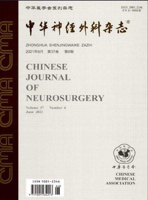
Chinese Journal of Neurosurgery is one of the series of journals organized by the Chinese Medical Association under the supervision of the China Association for Science and Technology. The journal is aimed at neurosurgeons and related researchers, and reports on the leading scientific research results and clinical experience in the field of neurosurgery, as well as the basic theoretical research closely related to neurosurgery.Chinese Journal of Neurosurgery has been included in many famous domestic search organizations, such as China Knowledge Resources Database, China Biomedical Journal Citation Database, Chinese Biomedical Journal Literature Database, China Science Citation Database, China Biomedical Literature Database, China Science and Technology Paper Citation Statistical Analysis Database, and China Science and Technology Journal Full Text Database, Wanfang Data Database of Medical Journals, etc.