Ioannis D. Gkegkes, Dimitrios I. Kapetanakis, Christos Iavazzo, Apostolos P. Stamatiadis
{"title":"一种不寻常的肛周溃疡","authors":"Ioannis D. Gkegkes, Dimitrios I. Kapetanakis, Christos Iavazzo, Apostolos P. Stamatiadis","doi":"10.1002/aid2.13364","DOIUrl":null,"url":null,"abstract":"<p>A 33-year-old male presented with a 14-day history of anal pain, mucus discharge and the sensation of incomplete evacuation after defecation. The patient's past medical history was significant for HIV infection, for which he was under treatment with elvitegravir/cobicistat/emtricitabine/tenofovir disoproxil fumarate (Stilbild®) daily. In addition, the patient was referred for unprotected anal intercourse 5 weeks prior to the onset of the symptoms.</p><p>The physical exam was significant for a perianal ulcer (Figure 1). On anoscopy of the anal canal, there was extensive inflammation of the anal mucosa. A three-dimensional (3D) endoanal ultrasound was also performed and did not show any evidence of any anal or perianal abscess. Bilateral inguinal lymph nodes were not enlarged. Furthermore, an anal ulcer swab was taken. Both Treponema pallidum hemagglutination assay (TPHA; index 15.4, positive: >1.1) and Venereal Disease Research Laboratory test (VDRL) were positive. The sample was processed with the use of polymerase chain reaction (PCR), which identified a <i>Treponema pallidum</i> infection. Primary perianal syphilis is often presented as a solitary, firm red papulae on the genital area which may progress to an ulcer with a well-defined margin and an indurated base. Patient received benzathine penicillin G 2.4 × 10<sup>6</sup> units in one intramuscular injection. The lesion was resolved within 5 weeks.</p><p>Worldwide, syphilis is considered a re-emerging public health problem.<span><sup>1</sup></span> Symptoms, such as anal pain, pus at the anal canal, signs of systematic involvement, and tenesmus should make clinicians suspicious of an anal sexually transmitted infection (STI). Men who have sex with men (MSM) have usually more sexual partners than heterosexual men, while they tend to have more simultaneous partners.<span><sup>2</sup></span> Moreover, in the last decades, HIV is no longer perceived to be a fatal disease, due to the fact that antiretroviral therapy suppresses effectively, there is a decrease on the use of condoms.<span><sup>2</sup></span> In addition, the administration of pre-exposure prophylaxis (PrEP) encourages unprotected intercourse among HIV-uninfected individuals (with low prevalence of syphilis), increasing the risk of contracting syphilis from people living with HIV (with a higher prevalence of syphilis).<span><sup>2</sup></span></p><p>The differential diagnosis of a perianal ulcer also includes perianal tuberculosis (Tb). Perianal Tb is a rare form of extrapulmonary tuberculosis and can be the initial manifestation of Tb.<span><sup>3</sup></span> Both Ziehl-Neelsen staining and culture, in addition to histopathological examination, are essential for achieving the correct diagnosis and to start the appropriate treatment.</p><p>Herpes simplex virus (HSV) infection should also be considered in the presence of perianal lesions.<span><sup>4</sup></span> HSV type 2 is the most common cause of genital and perianal ulcers. Patients typically present painful grouped vesicles, followed by painful superficial ulcers. The viral infection is easily diagnosed by cell culture, PCR testing, serology, and direct fluorescent antibody testing.</p><p>Chancroid is another STI that may present with painful sores on the genitalia.<span><sup>3</sup></span> Chancroid is a bacterial infection caused by <i>Haemophilus ducreyi</i>. The ulcer presents a painful serpiginous border and a friable base covered with a necrotic, often purulent exudate. Chancroid is diagnosed by a culture of the ulcer. In the scenario of a perianal ulcer, the probability of lymphogranuloma venereum (LV) should also be considered.<span><sup>3</sup></span> The etiological agent of LV is <i>Chlamydia trachomatis</i>, and it usually presents with a small, shallow, painless papule or ulcer. Additionally, LV is diagnosed by microbiological culture of the ulcers.</p><p>Finally, Adamantiades-Behcet's disease also enters the differential diagnosis. It is a chronic, systemic inflammatory disease of unknown etiology.<span><sup>5</sup></span> Genital and perianal ulcers are present in 70% to 90% of cases, along with aphthous oral ulcers.</p><p>The authors declare no conflicts of interest.</p><p>Written informed consent was obtained from the patient.</p>","PeriodicalId":7278,"journal":{"name":"Advances in Digestive Medicine","volume":"10 4","pages":"261-262"},"PeriodicalIF":0.4000,"publicationDate":"2023-07-18","publicationTypes":"Journal Article","fieldsOfStudy":null,"isOpenAccess":false,"openAccessPdf":"https://onlinelibrary.wiley.com/doi/epdf/10.1002/aid2.13364","citationCount":"0","resultStr":"{\"title\":\"An unusual perianal ulcer\",\"authors\":\"Ioannis D. Gkegkes, Dimitrios I. Kapetanakis, Christos Iavazzo, Apostolos P. Stamatiadis\",\"doi\":\"10.1002/aid2.13364\",\"DOIUrl\":null,\"url\":null,\"abstract\":\"<p>A 33-year-old male presented with a 14-day history of anal pain, mucus discharge and the sensation of incomplete evacuation after defecation. The patient's past medical history was significant for HIV infection, for which he was under treatment with elvitegravir/cobicistat/emtricitabine/tenofovir disoproxil fumarate (Stilbild®) daily. In addition, the patient was referred for unprotected anal intercourse 5 weeks prior to the onset of the symptoms.</p><p>The physical exam was significant for a perianal ulcer (Figure 1). On anoscopy of the anal canal, there was extensive inflammation of the anal mucosa. A three-dimensional (3D) endoanal ultrasound was also performed and did not show any evidence of any anal or perianal abscess. Bilateral inguinal lymph nodes were not enlarged. Furthermore, an anal ulcer swab was taken. Both Treponema pallidum hemagglutination assay (TPHA; index 15.4, positive: >1.1) and Venereal Disease Research Laboratory test (VDRL) were positive. The sample was processed with the use of polymerase chain reaction (PCR), which identified a <i>Treponema pallidum</i> infection. Primary perianal syphilis is often presented as a solitary, firm red papulae on the genital area which may progress to an ulcer with a well-defined margin and an indurated base. Patient received benzathine penicillin G 2.4 × 10<sup>6</sup> units in one intramuscular injection. The lesion was resolved within 5 weeks.</p><p>Worldwide, syphilis is considered a re-emerging public health problem.<span><sup>1</sup></span> Symptoms, such as anal pain, pus at the anal canal, signs of systematic involvement, and tenesmus should make clinicians suspicious of an anal sexually transmitted infection (STI). Men who have sex with men (MSM) have usually more sexual partners than heterosexual men, while they tend to have more simultaneous partners.<span><sup>2</sup></span> Moreover, in the last decades, HIV is no longer perceived to be a fatal disease, due to the fact that antiretroviral therapy suppresses effectively, there is a decrease on the use of condoms.<span><sup>2</sup></span> In addition, the administration of pre-exposure prophylaxis (PrEP) encourages unprotected intercourse among HIV-uninfected individuals (with low prevalence of syphilis), increasing the risk of contracting syphilis from people living with HIV (with a higher prevalence of syphilis).<span><sup>2</sup></span></p><p>The differential diagnosis of a perianal ulcer also includes perianal tuberculosis (Tb). Perianal Tb is a rare form of extrapulmonary tuberculosis and can be the initial manifestation of Tb.<span><sup>3</sup></span> Both Ziehl-Neelsen staining and culture, in addition to histopathological examination, are essential for achieving the correct diagnosis and to start the appropriate treatment.</p><p>Herpes simplex virus (HSV) infection should also be considered in the presence of perianal lesions.<span><sup>4</sup></span> HSV type 2 is the most common cause of genital and perianal ulcers. Patients typically present painful grouped vesicles, followed by painful superficial ulcers. The viral infection is easily diagnosed by cell culture, PCR testing, serology, and direct fluorescent antibody testing.</p><p>Chancroid is another STI that may present with painful sores on the genitalia.<span><sup>3</sup></span> Chancroid is a bacterial infection caused by <i>Haemophilus ducreyi</i>. The ulcer presents a painful serpiginous border and a friable base covered with a necrotic, often purulent exudate. Chancroid is diagnosed by a culture of the ulcer. In the scenario of a perianal ulcer, the probability of lymphogranuloma venereum (LV) should also be considered.<span><sup>3</sup></span> The etiological agent of LV is <i>Chlamydia trachomatis</i>, and it usually presents with a small, shallow, painless papule or ulcer. Additionally, LV is diagnosed by microbiological culture of the ulcers.</p><p>Finally, Adamantiades-Behcet's disease also enters the differential diagnosis. It is a chronic, systemic inflammatory disease of unknown etiology.<span><sup>5</sup></span> Genital and perianal ulcers are present in 70% to 90% of cases, along with aphthous oral ulcers.</p><p>The authors declare no conflicts of interest.</p><p>Written informed consent was obtained from the patient.</p>\",\"PeriodicalId\":7278,\"journal\":{\"name\":\"Advances in Digestive Medicine\",\"volume\":\"10 4\",\"pages\":\"261-262\"},\"PeriodicalIF\":0.4000,\"publicationDate\":\"2023-07-18\",\"publicationTypes\":\"Journal Article\",\"fieldsOfStudy\":null,\"isOpenAccess\":false,\"openAccessPdf\":\"https://onlinelibrary.wiley.com/doi/epdf/10.1002/aid2.13364\",\"citationCount\":\"0\",\"resultStr\":null,\"platform\":\"Semanticscholar\",\"paperid\":null,\"PeriodicalName\":\"Advances in Digestive Medicine\",\"FirstCategoryId\":\"1085\",\"ListUrlMain\":\"https://onlinelibrary.wiley.com/doi/10.1002/aid2.13364\",\"RegionNum\":0,\"RegionCategory\":null,\"ArticlePicture\":[],\"TitleCN\":null,\"AbstractTextCN\":null,\"PMCID\":null,\"EPubDate\":\"\",\"PubModel\":\"\",\"JCR\":\"Q4\",\"JCRName\":\"GASTROENTEROLOGY & HEPATOLOGY\",\"Score\":null,\"Total\":0}","platform":"Semanticscholar","paperid":null,"PeriodicalName":"Advances in Digestive Medicine","FirstCategoryId":"1085","ListUrlMain":"https://onlinelibrary.wiley.com/doi/10.1002/aid2.13364","RegionNum":0,"RegionCategory":null,"ArticlePicture":[],"TitleCN":null,"AbstractTextCN":null,"PMCID":null,"EPubDate":"","PubModel":"","JCR":"Q4","JCRName":"GASTROENTEROLOGY & HEPATOLOGY","Score":null,"Total":0}
A 33-year-old male presented with a 14-day history of anal pain, mucus discharge and the sensation of incomplete evacuation after defecation. The patient's past medical history was significant for HIV infection, for which he was under treatment with elvitegravir/cobicistat/emtricitabine/tenofovir disoproxil fumarate (Stilbild®) daily. In addition, the patient was referred for unprotected anal intercourse 5 weeks prior to the onset of the symptoms.
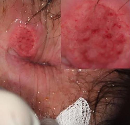
The physical exam was significant for a perianal ulcer (Figure 1). On anoscopy of the anal canal, there was extensive inflammation of the anal mucosa. A three-dimensional (3D) endoanal ultrasound was also performed and did not show any evidence of any anal or perianal abscess. Bilateral inguinal lymph nodes were not enlarged. Furthermore, an anal ulcer swab was taken. Both Treponema pallidum hemagglutination assay (TPHA; index 15.4, positive: >1.1) and Venereal Disease Research Laboratory test (VDRL) were positive. The sample was processed with the use of polymerase chain reaction (PCR), which identified a Treponema pallidum infection. Primary perianal syphilis is often presented as a solitary, firm red papulae on the genital area which may progress to an ulcer with a well-defined margin and an indurated base. Patient received benzathine penicillin G 2.4 × 10⁶ units in one intramuscular injection. The lesion was resolved within 5 weeks.
Worldwide, syphilis is considered a re-emerging public health problem.1 Symptoms, such as anal pain, pus at the anal canal, signs of systematic involvement, and tenesmus should make clinicians suspicious of an anal sexually transmitted infection (STI). Men who have sex with men (MSM) have usually more sexual partners than heterosexual men, while they tend to have more simultaneous partners.2 Moreover, in the last decades, HIV is no longer perceived to be a fatal disease, due to the fact that antiretroviral therapy suppresses effectively, there is a decrease on the use of condoms.2 In addition, the administration of pre-exposure prophylaxis (PrEP) encourages unprotected intercourse among HIV-uninfected individuals (with low prevalence of syphilis), increasing the risk of contracting syphilis from people living with HIV (with a higher prevalence of syphilis).2
The differential diagnosis of a perianal ulcer also includes perianal tuberculosis (Tb). Perianal Tb is a rare form of extrapulmonary tuberculosis and can be the initial manifestation of Tb.3 Both Ziehl-Neelsen staining and culture, in addition to histopathological examination, are essential for achieving the correct diagnosis and to start the appropriate treatment.
Herpes simplex virus (HSV) infection should also be considered in the presence of perianal lesions.4 HSV type 2 is the most common cause of genital and perianal ulcers. Patients typically present painful grouped vesicles, followed by painful superficial ulcers. The viral infection is easily diagnosed by cell culture, PCR testing, serology, and direct fluorescent antibody testing.
Chancroid is another STI that may present with painful sores on the genitalia.3 Chancroid is a bacterial infection caused by Haemophilus ducreyi. The ulcer presents a painful serpiginous border and a friable base covered with a necrotic, often purulent exudate. Chancroid is diagnosed by a culture of the ulcer. In the scenario of a perianal ulcer, the probability of lymphogranuloma venereum (LV) should also be considered.3 The etiological agent of LV is Chlamydia trachomatis, and it usually presents with a small, shallow, painless papule or ulcer. Additionally, LV is diagnosed by microbiological culture of the ulcers.
Finally, Adamantiades-Behcet's disease also enters the differential diagnosis. It is a chronic, systemic inflammatory disease of unknown etiology.5 Genital and perianal ulcers are present in 70% to 90% of cases, along with aphthous oral ulcers.
The authors declare no conflicts of interest.
Written informed consent was obtained from the patient.
期刊介绍:
Advances in Digestive Medicine is the official peer-reviewed journal of GEST, DEST and TASL. Missions of AIDM are to enhance the quality of patient care, to promote researches in gastroenterology, endoscopy and hepatology related fields, and to develop platforms for digestive science. Specific areas of interest are included, but not limited to: • Acid-related disease • Small intestinal disease • Digestive cancer • Diagnostic & therapeutic endoscopy • Enteral nutrition • Innovation in endoscopic technology • Functional GI • Hepatitis • GI images • Liver cirrhosis • Gut hormone • NASH • Helicobacter pylori • Cancer screening • IBD • Laparoscopic surgery • Infectious disease of digestive tract • Genetics and metabolic disorder • Microbiota • Regenerative medicine • Pancreaticobiliary disease • Guideline & consensus.

 求助内容:
求助内容: 应助结果提醒方式:


