局部晚期声门上鳞癌淋巴结转移模式及危险因素分析

Yi Xu, Ye Zhang, Shaoyan Liu, Xiaolei Wang, Xiao-dong Huang, Y. Qu, Kai Wang, R. Wu, Xue-song Chen, Qingfeng Liu, Shiran Sun, Xi Luo, Jingwei Luo, Li Gao, Guo-zhen Xu, J. Yi
{"title":"局部晚期声门上鳞癌淋巴结转移模式及危险因素分析","authors":"Yi Xu, Ye Zhang, Shaoyan Liu, Xiaolei Wang, Xiao-dong Huang, Y. Qu, Kai Wang, R. Wu, Xue-song Chen, Qingfeng Liu, Shiran Sun, Xi Luo, Jingwei Luo, Li Gao, Guo-zhen Xu, J. Yi","doi":"10.3760/CMA.J.ISSN.1004-4221.2020.01.002","DOIUrl":null,"url":null,"abstract":"Objective \nTo investigate the pattern of lymph node metastasis (LNM) in patients with locally advanced (T3, T4) laryngeal squamous cell carcinoma (LALSC) and provide reference for the delineation of clinical target volume. \n \n \nMethods \nClinical data of 272 patients with LALSC treated in our hospital from 2000 to 2017 were retrospectively analyzed. All patients underwent bilateral neck dissection (at least level Ⅱ-Ⅳ). The LNM ratio of each node level was calculated. The risk factors of LNM were identified by univariate and multivariate logistic regression analyses. \n \n \nResults \nLNM was found in 156 of 272 patients (57.1%). According to the location of primary lesions, all patients were divided into group A (n=72; unilateral without midline involvement), group B (n=86; unilateral with midline involvement) and group C (n=114; giant or central). In group A, the LNM ratio at ipsilateral level Ⅱ, Ⅲ and Ⅳ was 36.3%, 26.4% and 6.9%, whereas 13.9%, 8.3% and 1.4% at the contralateral level, respectively. In group B, the LNM ratio at ipsilateral level Ⅱ, Ⅲ and IV was 1.9%, 29.1% and 11.6%, whereas 18.6%, 14.0% and 1.2% at the contralateral level, respectively. In group C, the LNM ratio at the left neck level Ⅱ, Ⅲ and Ⅳ was 24.6%, 23.7% and 2.6%, whereas 21.9%, 26.3% and 6.1% at the right neck, respectively. Bilateral LNM ratio did not significantly differ between group A and group B/C (15.3%, 25.0%, P=0.093). Ipsilateral level Ⅲ metastasis (OR=2.929, 95%CI 1.041-8.245, P=0.042) and clinical N stage (OR=0.082, 95%CI 0.018-0.373, P=0.001) were associated with contralateral LNM. Ipsilateral level Ⅱ(P=0.043) or Ⅲ(P=0.009) metastasis were risk factors of the ipsilateral level Ⅳ metastasis. \n \n \nConclusions \nNeck levels Ⅱ and Ⅲ are the high-risk LNM regions, whereaslevels Ⅳ and V are the low-risk areas. Ipsilateral level Ⅱ or Ⅲ metastases are the risk factors of ipsilateral level Ⅳ and contralateral cervical LNM. Contralateral neck LNM rarely occurs in cN0 stage patients. \n \n \nKey words: \nLocally advanced laryngeal cancer; Squamous cell carcinoma; Lymph node metastasis; Risk factor","PeriodicalId":10288,"journal":{"name":"中华放射肿瘤学杂志","volume":"29 1","pages":"6-10"},"PeriodicalIF":0.0000,"publicationDate":"2020-01-15","publicationTypes":"Journal Article","fieldsOfStudy":null,"isOpenAccess":false,"openAccessPdf":"","citationCount":"0","resultStr":"{\"title\":\"Patterns and risk factors of lymph node metastasis in locally advanced supraglottic squamous cell carcinoma\",\"authors\":\"Yi Xu, Ye Zhang, Shaoyan Liu, Xiaolei Wang, Xiao-dong Huang, Y. Qu, Kai Wang, R. Wu, Xue-song Chen, Qingfeng Liu, Shiran Sun, Xi Luo, Jingwei Luo, Li Gao, Guo-zhen Xu, J. Yi\",\"doi\":\"10.3760/CMA.J.ISSN.1004-4221.2020.01.002\",\"DOIUrl\":null,\"url\":null,\"abstract\":\"Objective \\nTo investigate the pattern of lymph node metastasis (LNM) in patients with locally advanced (T3, T4) laryngeal squamous cell carcinoma (LALSC) and provide reference for the delineation of clinical target volume. \\n \\n \\nMethods \\nClinical data of 272 patients with LALSC treated in our hospital from 2000 to 2017 were retrospectively analyzed. All patients underwent bilateral neck dissection (at least level Ⅱ-Ⅳ). The LNM ratio of each node level was calculated. The risk factors of LNM were identified by univariate and multivariate logistic regression analyses. \\n \\n \\nResults \\nLNM was found in 156 of 272 patients (57.1%). According to the location of primary lesions, all patients were divided into group A (n=72; unilateral without midline involvement), group B (n=86; unilateral with midline involvement) and group C (n=114; giant or central). In group A, the LNM ratio at ipsilateral level Ⅱ, Ⅲ and Ⅳ was 36.3%, 26.4% and 6.9%, whereas 13.9%, 8.3% and 1.4% at the contralateral level, respectively. In group B, the LNM ratio at ipsilateral level Ⅱ, Ⅲ and IV was 1.9%, 29.1% and 11.6%, whereas 18.6%, 14.0% and 1.2% at the contralateral level, respectively. In group C, the LNM ratio at the left neck level Ⅱ, Ⅲ and Ⅳ was 24.6%, 23.7% and 2.6%, whereas 21.9%, 26.3% and 6.1% at the right neck, respectively. Bilateral LNM ratio did not significantly differ between group A and group B/C (15.3%, 25.0%, P=0.093). Ipsilateral level Ⅲ metastasis (OR=2.929, 95%CI 1.041-8.245, P=0.042) and clinical N stage (OR=0.082, 95%CI 0.018-0.373, P=0.001) were associated with contralateral LNM. Ipsilateral level Ⅱ(P=0.043) or Ⅲ(P=0.009) metastasis were risk factors of the ipsilateral level Ⅳ metastasis. \\n \\n \\nConclusions \\nNeck levels Ⅱ and Ⅲ are the high-risk LNM regions, whereaslevels Ⅳ and V are the low-risk areas. Ipsilateral level Ⅱ or Ⅲ metastases are the risk factors of ipsilateral level Ⅳ and contralateral cervical LNM. Contralateral neck LNM rarely occurs in cN0 stage patients. \\n \\n \\nKey words: \\nLocally advanced laryngeal cancer; Squamous cell carcinoma; Lymph node metastasis; Risk factor\",\"PeriodicalId\":10288,\"journal\":{\"name\":\"中华放射肿瘤学杂志\",\"volume\":\"29 1\",\"pages\":\"6-10\"},\"PeriodicalIF\":0.0000,\"publicationDate\":\"2020-01-15\",\"publicationTypes\":\"Journal Article\",\"fieldsOfStudy\":null,\"isOpenAccess\":false,\"openAccessPdf\":\"\",\"citationCount\":\"0\",\"resultStr\":null,\"platform\":\"Semanticscholar\",\"paperid\":null,\"PeriodicalName\":\"中华放射肿瘤学杂志\",\"FirstCategoryId\":\"3\",\"ListUrlMain\":\"https://doi.org/10.3760/CMA.J.ISSN.1004-4221.2020.01.002\",\"RegionNum\":0,\"RegionCategory\":null,\"ArticlePicture\":[],\"TitleCN\":null,\"AbstractTextCN\":null,\"PMCID\":null,\"EPubDate\":\"\",\"PubModel\":\"\",\"JCR\":\"\",\"JCRName\":\"\",\"Score\":null,\"Total\":0}","platform":"Semanticscholar","paperid":null,"PeriodicalName":"中华放射肿瘤学杂志","FirstCategoryId":"3","ListUrlMain":"https://doi.org/10.3760/CMA.J.ISSN.1004-4221.2020.01.002","RegionNum":0,"RegionCategory":null,"ArticlePicture":[],"TitleCN":null,"AbstractTextCN":null,"PMCID":null,"EPubDate":"","PubModel":"","JCR":"","JCRName":"","Score":null,"Total":0}
引用次数: 0

摘要

目的探讨局部晚期(T3、T4)喉鳞癌(LALSC)患者的淋巴结转移(LNM)模式,为临床靶体积的划定提供参考。方法回顾性分析我院2000 ~ 2017年收治的272例LALSC患者的临床资料。所有患者均行双侧颈部清扫术(至少Ⅱ-Ⅳ段)。计算各节点级别的LNM比率。通过单因素和多因素logistic回归分析确定LNM的危险因素。结果272例患者中有156例出现LNM,占57.1%。根据原发病灶的位置,将所有患者分为A组(n=72;单侧无中线受累),B组(n=86;单侧中线受累)和C组(n=114;巨型或中央)。A组同侧Ⅱ、Ⅲ和Ⅳ的LNM比例分别为36.3%、26.4%和6.9%,对侧分别为13.9%、8.3%和1.4%。B组同侧Ⅱ、Ⅲ和IV的LNM率分别为1.9%、29.1%和11.6%,对侧LNM率分别为18.6%、14.0%和1.2%。C组左颈部水平Ⅱ、Ⅲ和Ⅳ的LNM比例分别为24.6%、23.7%和2.6%,右颈部水平分别为21.9%、26.3%和6.1%。A组与B/C组双侧LNM比例差异无统计学意义(15.3%,25.0%,P=0.093)。同侧水平Ⅲ转移(OR=2.929, 95%CI 1.041 ~ 8.245, P=0.042)和临床N分期(OR=0.082, 95%CI 0.018 ~ 0.373, P=0.001)与对侧LNM相关。同侧水平Ⅱ(P=0.043)或Ⅲ(P=0.009)转移是同侧水平Ⅳ转移的危险因素。结论颈部级Ⅱ、Ⅲ为LNM的高危区,Ⅳ、V级为LNM的低危区。同侧水平Ⅱ或Ⅲ转移是同侧水平Ⅳ和对侧宫颈LNM的危险因素。对侧颈部LNM很少发生在cN0期患者。关键词:局部晚期喉癌;鳞状细胞癌;淋巴结转移;风险因素
本文章由计算机程序翻译,如有差异,请以英文原文为准。
Patterns and risk factors of lymph node metastasis in locally advanced supraglottic squamous cell carcinoma
Objective To investigate the pattern of lymph node metastasis (LNM) in patients with locally advanced (T3, T4) laryngeal squamous cell carcinoma (LALSC) and provide reference for the delineation of clinical target volume. Methods Clinical data of 272 patients with LALSC treated in our hospital from 2000 to 2017 were retrospectively analyzed. All patients underwent bilateral neck dissection (at least level Ⅱ-Ⅳ). The LNM ratio of each node level was calculated. The risk factors of LNM were identified by univariate and multivariate logistic regression analyses. Results LNM was found in 156 of 272 patients (57.1%). According to the location of primary lesions, all patients were divided into group A (n=72; unilateral without midline involvement), group B (n=86; unilateral with midline involvement) and group C (n=114; giant or central). In group A, the LNM ratio at ipsilateral level Ⅱ, Ⅲ and Ⅳ was 36.3%, 26.4% and 6.9%, whereas 13.9%, 8.3% and 1.4% at the contralateral level, respectively. In group B, the LNM ratio at ipsilateral level Ⅱ, Ⅲ and IV was 1.9%, 29.1% and 11.6%, whereas 18.6%, 14.0% and 1.2% at the contralateral level, respectively. In group C, the LNM ratio at the left neck level Ⅱ, Ⅲ and Ⅳ was 24.6%, 23.7% and 2.6%, whereas 21.9%, 26.3% and 6.1% at the right neck, respectively. Bilateral LNM ratio did not significantly differ between group A and group B/C (15.3%, 25.0%, P=0.093). Ipsilateral level Ⅲ metastasis (OR=2.929, 95%CI 1.041-8.245, P=0.042) and clinical N stage (OR=0.082, 95%CI 0.018-0.373, P=0.001) were associated with contralateral LNM. Ipsilateral level Ⅱ(P=0.043) or Ⅲ(P=0.009) metastasis were risk factors of the ipsilateral level Ⅳ metastasis. Conclusions Neck levels Ⅱ and Ⅲ are the high-risk LNM regions, whereaslevels Ⅳ and V are the low-risk areas. Ipsilateral level Ⅱ or Ⅲ metastases are the risk factors of ipsilateral level Ⅳ and contralateral cervical LNM. Contralateral neck LNM rarely occurs in cN0 stage patients. Key words: Locally advanced laryngeal cancer; Squamous cell carcinoma; Lymph node metastasis; Risk factor
求助全文
通过发布文献求助,成功后即可免费获取论文全文。 去求助
来源期刊
自引率
0.00%
发文量
6375
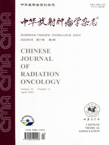
期刊介绍: The Chinese Journal of Radiation Oncology is a national academic journal sponsored by the Chinese Medical Association. It was founded in 1992 and the title was written by Chen Minzhang, the former Minister of Health. Its predecessor was the Chinese Journal of Radiation Oncology, which was founded in 1987. The journal is an authoritative journal in the field of radiation oncology in my country. It focuses on clinical tumor radiotherapy, tumor radiation physics, tumor radiation biology, and thermal therapy. Its main readers are middle and senior clinical doctors and scientific researchers. It is now a monthly journal with a large 16-page format and 80 pages of text. For many years, it has adhered to the principle of combining theory with practice and combining improvement with popularization. It now has columns such as monographs, head and neck tumors (monographs), chest tumors (monographs), abdominal tumors (monographs), physics, technology, biology (monographs), reviews, and investigations and research.
×
引用
GB/T 7714-2015
复制
MLA
复制
APA
复制
导出至
BibTeX EndNote RefMan NoteFirst NoteExpress
×
提示
您的信息不完整,为了账户安全,请先补充。
现在去补充
×
提示
您因"违规操作"
具体请查看互助需知
我知道了
×
提示
确定
请完成安全验证×
copy
已复制链接
快去分享给好友吧!
我知道了
右上角分享
点击右上角分享
0
联系我们:info@booksci.cn Book学术提供免费学术资源搜索服务,方便国内外学者检索中英文文献。致力于提供最便捷和优质的服务体验。 Copyright © 2023 布克学术 All rights reserved.
京ICP备2023020795号-1
ghs 京公网安备 11010802042870号
Book学术文献互助
Book学术文献互助群
群 号:481959085
Book学术官方微信