磁共振成像在胆囊结石伴胆囊壁弥漫性炎性增厚患者治疗途径选择中的指导作用

Q4 Medicine
Kun Yan, Z. Ding, Guoping Chen, Jianjun Zheng, Yinhua Jin, Bibo Hu, Bin Chen, Jingfeng Zhang
{"title":"磁共振成像在胆囊结石伴胆囊壁弥漫性炎性增厚患者治疗途径选择中的指导作用","authors":"Kun Yan, Z. Ding, Guoping Chen, Jianjun Zheng, Yinhua Jin, Bibo Hu, Bin Chen, Jingfeng Zhang","doi":"10.3760/CMA.J.ISSN.1007-8118.2019.12.005","DOIUrl":null,"url":null,"abstract":"Objective \nTo investigate the use of conventional MR imaging to guide treatment in patients with cholecystolithiasis and diffuse inflammatory thickening of gallbladder wall. \n \n \nMethods \nThe clinical data of patients who were treated in the Ningbo Huamei Hospital, University of the Chinese Academy of Sciences between January 2017 and January 2018 were analyzed. These patients were divided into two groups: patients with acute cholecystitis (n=139) and patients with viral hepatitis combined with cholecystolithiasis (n=67). Differences in the imaging signs in standardized upper abdominal contrast enhanced MRI examinations were retrospectively analyzed. \n \n \nResults \nThe imaging signs, including stone location, continuity of gallbladder mucosa, exudation in peri-gallbladder space, edema of intrahepatic portal area showed significant differences between the two groups (all P<0.05). On stratification analysis, the type of thickened gallbladder wall, background of liver parenchyma and extent of edema in intrahepatic catchment area also showed significant differences (all P<0.05). The imaging signs, including non-gallbladder neck ductal stones, concentric thickening of gallbladder wall, continuous mucous membrane in gallbladder and no peri-gallbladder space exudation but diffuse edema of intrahepatic catchment area supported the diagnosis of viral hepatitis combined with gallstones. The imaging signs, including discontinuity of gallbladder mucosa, exudation of peri-gallbladder space, diffuse edema of gallbladder wall without a cirrhotic background and edema in intrahepatic portal area supported the diagnosis of acute calculous cholecystitis of gallbladder. \n \n \nConclusions \nRoutine upper abdominal contrast enhanced MRI plays an important role in demonstrating the underlying cause of gallbladder wall diffuse edema thickening in patients with gallstones. It provides an important reference for the choice of clinical treatment pathway. \n \n \nKey words: \nCholecystolithiasis; Clinical protocols; Cholecystitis; Hepatitis B; Magnetic resonance imaging","PeriodicalId":10021,"journal":{"name":"中华肝胆外科杂志","volume":"25 1","pages":"899-904"},"PeriodicalIF":0.0000,"publicationDate":"2019-12-28","publicationTypes":"Journal Article","fieldsOfStudy":null,"isOpenAccess":false,"openAccessPdf":"","citationCount":"0","resultStr":"{\"title\":\"Magnetic resonance imaging in guiding choice of treatment pathway in patients with cholecystolithiasis and diffuse inflammatory thickening of gallbladder wall\",\"authors\":\"Kun Yan, Z. Ding, Guoping Chen, Jianjun Zheng, Yinhua Jin, Bibo Hu, Bin Chen, Jingfeng Zhang\",\"doi\":\"10.3760/CMA.J.ISSN.1007-8118.2019.12.005\",\"DOIUrl\":null,\"url\":null,\"abstract\":\"Objective \\nTo investigate the use of conventional MR imaging to guide treatment in patients with cholecystolithiasis and diffuse inflammatory thickening of gallbladder wall. \\n \\n \\nMethods \\nThe clinical data of patients who were treated in the Ningbo Huamei Hospital, University of the Chinese Academy of Sciences between January 2017 and January 2018 were analyzed. These patients were divided into two groups: patients with acute cholecystitis (n=139) and patients with viral hepatitis combined with cholecystolithiasis (n=67). Differences in the imaging signs in standardized upper abdominal contrast enhanced MRI examinations were retrospectively analyzed. \\n \\n \\nResults \\nThe imaging signs, including stone location, continuity of gallbladder mucosa, exudation in peri-gallbladder space, edema of intrahepatic portal area showed significant differences between the two groups (all P<0.05). On stratification analysis, the type of thickened gallbladder wall, background of liver parenchyma and extent of edema in intrahepatic catchment area also showed significant differences (all P<0.05). The imaging signs, including non-gallbladder neck ductal stones, concentric thickening of gallbladder wall, continuous mucous membrane in gallbladder and no peri-gallbladder space exudation but diffuse edema of intrahepatic catchment area supported the diagnosis of viral hepatitis combined with gallstones. The imaging signs, including discontinuity of gallbladder mucosa, exudation of peri-gallbladder space, diffuse edema of gallbladder wall without a cirrhotic background and edema in intrahepatic portal area supported the diagnosis of acute calculous cholecystitis of gallbladder. \\n \\n \\nConclusions \\nRoutine upper abdominal contrast enhanced MRI plays an important role in demonstrating the underlying cause of gallbladder wall diffuse edema thickening in patients with gallstones. It provides an important reference for the choice of clinical treatment pathway. \\n \\n \\nKey words: \\nCholecystolithiasis; Clinical protocols; Cholecystitis; Hepatitis B; Magnetic resonance imaging\",\"PeriodicalId\":10021,\"journal\":{\"name\":\"中华肝胆外科杂志\",\"volume\":\"25 1\",\"pages\":\"899-904\"},\"PeriodicalIF\":0.0000,\"publicationDate\":\"2019-12-28\",\"publicationTypes\":\"Journal Article\",\"fieldsOfStudy\":null,\"isOpenAccess\":false,\"openAccessPdf\":\"\",\"citationCount\":\"0\",\"resultStr\":null,\"platform\":\"Semanticscholar\",\"paperid\":null,\"PeriodicalName\":\"中华肝胆外科杂志\",\"FirstCategoryId\":\"3\",\"ListUrlMain\":\"https://doi.org/10.3760/CMA.J.ISSN.1007-8118.2019.12.005\",\"RegionNum\":0,\"RegionCategory\":null,\"ArticlePicture\":[],\"TitleCN\":null,\"AbstractTextCN\":null,\"PMCID\":null,\"EPubDate\":\"\",\"PubModel\":\"\",\"JCR\":\"Q4\",\"JCRName\":\"Medicine\",\"Score\":null,\"Total\":0}","platform":"Semanticscholar","paperid":null,"PeriodicalName":"中华肝胆外科杂志","FirstCategoryId":"3","ListUrlMain":"https://doi.org/10.3760/CMA.J.ISSN.1007-8118.2019.12.005","RegionNum":0,"RegionCategory":null,"ArticlePicture":[],"TitleCN":null,"AbstractTextCN":null,"PMCID":null,"EPubDate":"","PubModel":"","JCR":"Q4","JCRName":"Medicine","Score":null,"Total":0}
引用次数: 0

摘要

目的探讨应用常规磁共振成像技术指导胆囊结石合并胆囊壁弥漫性炎症性增厚患者的治疗。方法分析2017年1月至2018年1月在中国科学院大学宁波华美医院就诊的患者的临床资料。将这些患者分为两组:急性胆囊炎患者(n=139)和病毒性肝炎合并胆囊结石患者(n=67)。回顾性分析标准化上腹造影增强MRI检查中影像学征象的差异。结果两组在结石位置、胆囊黏膜连续性、胆囊周围渗出、肝内门静脉水肿等影像学表现上有显著性差异(均P<0.05),肝实质背景和肝内汇水区水肿程度也有显著性差异(均P<0.05),胆囊黏膜连续,无胆囊周围间隙渗出,肝内汇水区弥漫性水肿,支持病毒性肝炎合并胆结石的诊断。胆囊黏膜不连续、胆囊周围间隙渗出、无肝硬化背景的胆囊壁弥漫性水肿、肝内门区水肿等影像学征象支持了对胆囊急性结石性胆囊炎的诊断。结论常规上腹部对比增强MRI在揭示胆囊结石患者胆囊壁弥漫性水肿增厚的根本原因方面起着重要作用。为临床治疗路径的选择提供了重要参考。关键词:胆囊结石;临床方案;胆囊炎;乙型肝炎;磁共振成像
本文章由计算机程序翻译,如有差异,请以英文原文为准。
Magnetic resonance imaging in guiding choice of treatment pathway in patients with cholecystolithiasis and diffuse inflammatory thickening of gallbladder wall
Objective To investigate the use of conventional MR imaging to guide treatment in patients with cholecystolithiasis and diffuse inflammatory thickening of gallbladder wall. Methods The clinical data of patients who were treated in the Ningbo Huamei Hospital, University of the Chinese Academy of Sciences between January 2017 and January 2018 were analyzed. These patients were divided into two groups: patients with acute cholecystitis (n=139) and patients with viral hepatitis combined with cholecystolithiasis (n=67). Differences in the imaging signs in standardized upper abdominal contrast enhanced MRI examinations were retrospectively analyzed. Results The imaging signs, including stone location, continuity of gallbladder mucosa, exudation in peri-gallbladder space, edema of intrahepatic portal area showed significant differences between the two groups (all P<0.05). On stratification analysis, the type of thickened gallbladder wall, background of liver parenchyma and extent of edema in intrahepatic catchment area also showed significant differences (all P<0.05). The imaging signs, including non-gallbladder neck ductal stones, concentric thickening of gallbladder wall, continuous mucous membrane in gallbladder and no peri-gallbladder space exudation but diffuse edema of intrahepatic catchment area supported the diagnosis of viral hepatitis combined with gallstones. The imaging signs, including discontinuity of gallbladder mucosa, exudation of peri-gallbladder space, diffuse edema of gallbladder wall without a cirrhotic background and edema in intrahepatic portal area supported the diagnosis of acute calculous cholecystitis of gallbladder. Conclusions Routine upper abdominal contrast enhanced MRI plays an important role in demonstrating the underlying cause of gallbladder wall diffuse edema thickening in patients with gallstones. It provides an important reference for the choice of clinical treatment pathway. Key words: Cholecystolithiasis; Clinical protocols; Cholecystitis; Hepatitis B; Magnetic resonance imaging
求助全文
通过发布文献求助,成功后即可免费获取论文全文。 去求助
来源期刊
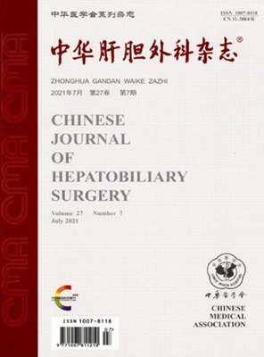
中华肝胆外科杂志
中华肝胆外科杂志 Medicine-Gastroenterology
CiteScore
0.20
自引率
0.00%
发文量
7101
期刊介绍: Chinese Journal of Hepatobiliary Surgery is an academic journal organized by the Chinese Medical Association and supervised by the China Association for Science and Technology, founded in 1995. The journal has the following columns: review, hot spotlight, academic thinking, thesis, experimental research, short thesis, case report, synthesis, etc. The journal has been recognized by Beida Journal (Chinese Journal of Humanities and Social Sciences). Chinese Journal of Hepatobiliary Surgery has been included in famous databases such as Peking University Journal (Chinese Journal of Humanities and Social Sciences), CSCD Source Journals of China Science Citation Database (with Extended Version) and so on, and it is one of the national key academic journals under the supervision of China Association for Science and Technology.
×
引用
GB/T 7714-2015
复制
MLA
复制
APA
复制
导出至
BibTeX EndNote RefMan NoteFirst NoteExpress
×
提示
您的信息不完整,为了账户安全,请先补充。
现在去补充
×
提示
您因"违规操作"
具体请查看互助需知
我知道了
×
提示
确定
请完成安全验证×
copy
已复制链接
快去分享给好友吧!
我知道了
右上角分享
点击右上角分享
0
联系我们:info@booksci.cn Book学术提供免费学术资源搜索服务,方便国内外学者检索中英文文献。致力于提供最便捷和优质的服务体验。 Copyright © 2023 布克学术 All rights reserved.
京ICP备2023020795号-1
ghs 京公网安备 11010802042870号
Book学术文献互助
Book学术文献互助群
群 号:481959085
Book学术官方微信