Sofía S Sánchez-Boluarte, Wilfor Aguirre-Quispe, Manuel Herrera Aramburú, William O Tatum, Walter De La Cruz Ramírez
{"title":"术中皮质电图上短暂的潜在的节律性放电预测手术切除后局灶性皮质发育不良的良好结果:一个病例报告","authors":"Sofía S Sánchez-Boluarte, Wilfor Aguirre-Quispe, Manuel Herrera Aramburú, William O Tatum, Walter De La Cruz Ramírez","doi":"10.1186/s42494-023-00131-6","DOIUrl":null,"url":null,"abstract":"<p><strong>Background: </strong>Focal cortical dysplasia (FCD) is a common cause of drug-resistant epilepsy. Electroencephalography (EEG) biomarkers that predict good postoperative outcomes are essential for identifying patients with focal epilepsies.</p><p><strong>Case presentation: </strong>We report the case of a 21-year-old female with seizure onset at the age of 9, characterized by left-hand dystonic posturing and impaired awareness, which evolved to bilateral tonic-clonic seizures, evaluated in a neurological referral center in Lima, Peru. During 6-h video-EEG, interictal EEG revealing focal brief potentially ictal rhythmic discharges (BIRDs) over the right frontal central region, lasting less than 10 s. The ictal features were characterized by low-voltage fast activity over the same area. Brain magnetic resonance imaging (MRI) demonstrated a focal lesion of focal cortical dysplasia type II in the right frontal lobe. The patient underwent a lesionectomy guided by electrocorticography, which showed continuous polyspikes. BIRDs showing a brief burst of spikes lasting longer than 0.5 s, were also identified on intraoperative electrocorticography (ECoG) and helped define the extent of resection. The patient obtained an Engel Outcome Class IA at 6 years of follow-up.</p><p><strong>Conclusions: </strong>The atypical BIRDs on ECoG can be used as a prognostic biomarker for prolonged seizure-freedom outcome in patients with epilepsy. Additional reports are needed in developing countries with and without brain MRI lesions to advance outpatient presurgical evaluations despite limited resources.</p>","PeriodicalId":33628,"journal":{"name":"Acta Epileptologica","volume":"5 1","pages":"18"},"PeriodicalIF":1.2000,"publicationDate":"2023-07-18","publicationTypes":"Journal Article","fieldsOfStudy":null,"isOpenAccess":false,"openAccessPdf":"https://www.ncbi.nlm.nih.gov/pmc/articles/PMC11960384/pdf/","citationCount":"0","resultStr":"{\"title\":\"Brief potentially ictal rhythmic discharges on intraoperative electrocorticography predict a good outcome of focal cortical dysplasia after surgical resection: a case report.\",\"authors\":\"Sofía S Sánchez-Boluarte, Wilfor Aguirre-Quispe, Manuel Herrera Aramburú, William O Tatum, Walter De La Cruz Ramírez\",\"doi\":\"10.1186/s42494-023-00131-6\",\"DOIUrl\":null,\"url\":null,\"abstract\":\"<p><strong>Background: </strong>Focal cortical dysplasia (FCD) is a common cause of drug-resistant epilepsy. Electroencephalography (EEG) biomarkers that predict good postoperative outcomes are essential for identifying patients with focal epilepsies.</p><p><strong>Case presentation: </strong>We report the case of a 21-year-old female with seizure onset at the age of 9, characterized by left-hand dystonic posturing and impaired awareness, which evolved to bilateral tonic-clonic seizures, evaluated in a neurological referral center in Lima, Peru. During 6-h video-EEG, interictal EEG revealing focal brief potentially ictal rhythmic discharges (BIRDs) over the right frontal central region, lasting less than 10 s. The ictal features were characterized by low-voltage fast activity over the same area. Brain magnetic resonance imaging (MRI) demonstrated a focal lesion of focal cortical dysplasia type II in the right frontal lobe. The patient underwent a lesionectomy guided by electrocorticography, which showed continuous polyspikes. BIRDs showing a brief burst of spikes lasting longer than 0.5 s, were also identified on intraoperative electrocorticography (ECoG) and helped define the extent of resection. The patient obtained an Engel Outcome Class IA at 6 years of follow-up.</p><p><strong>Conclusions: </strong>The atypical BIRDs on ECoG can be used as a prognostic biomarker for prolonged seizure-freedom outcome in patients with epilepsy. Additional reports are needed in developing countries with and without brain MRI lesions to advance outpatient presurgical evaluations despite limited resources.</p>\",\"PeriodicalId\":33628,\"journal\":{\"name\":\"Acta Epileptologica\",\"volume\":\"5 1\",\"pages\":\"18\"},\"PeriodicalIF\":1.2000,\"publicationDate\":\"2023-07-18\",\"publicationTypes\":\"Journal Article\",\"fieldsOfStudy\":null,\"isOpenAccess\":false,\"openAccessPdf\":\"https://www.ncbi.nlm.nih.gov/pmc/articles/PMC11960384/pdf/\",\"citationCount\":\"0\",\"resultStr\":null,\"platform\":\"Semanticscholar\",\"paperid\":null,\"PeriodicalName\":\"Acta Epileptologica\",\"FirstCategoryId\":\"1085\",\"ListUrlMain\":\"https://doi.org/10.1186/s42494-023-00131-6\",\"RegionNum\":0,\"RegionCategory\":null,\"ArticlePicture\":[],\"TitleCN\":null,\"AbstractTextCN\":null,\"PMCID\":null,\"EPubDate\":\"\",\"PubModel\":\"\",\"JCR\":\"Q4\",\"JCRName\":\"CLINICAL NEUROLOGY\",\"Score\":null,\"Total\":0}","platform":"Semanticscholar","paperid":null,"PeriodicalName":"Acta Epileptologica","FirstCategoryId":"1085","ListUrlMain":"https://doi.org/10.1186/s42494-023-00131-6","RegionNum":0,"RegionCategory":null,"ArticlePicture":[],"TitleCN":null,"AbstractTextCN":null,"PMCID":null,"EPubDate":"","PubModel":"","JCR":"Q4","JCRName":"CLINICAL NEUROLOGY","Score":null,"Total":0}
Brief potentially ictal rhythmic discharges on intraoperative electrocorticography predict a good outcome of focal cortical dysplasia after surgical resection: a case report.
Background: Focal cortical dysplasia (FCD) is a common cause of drug-resistant epilepsy. Electroencephalography (EEG) biomarkers that predict good postoperative outcomes are essential for identifying patients with focal epilepsies.
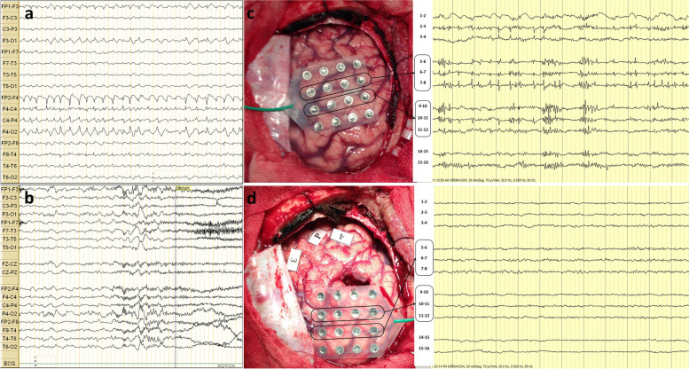
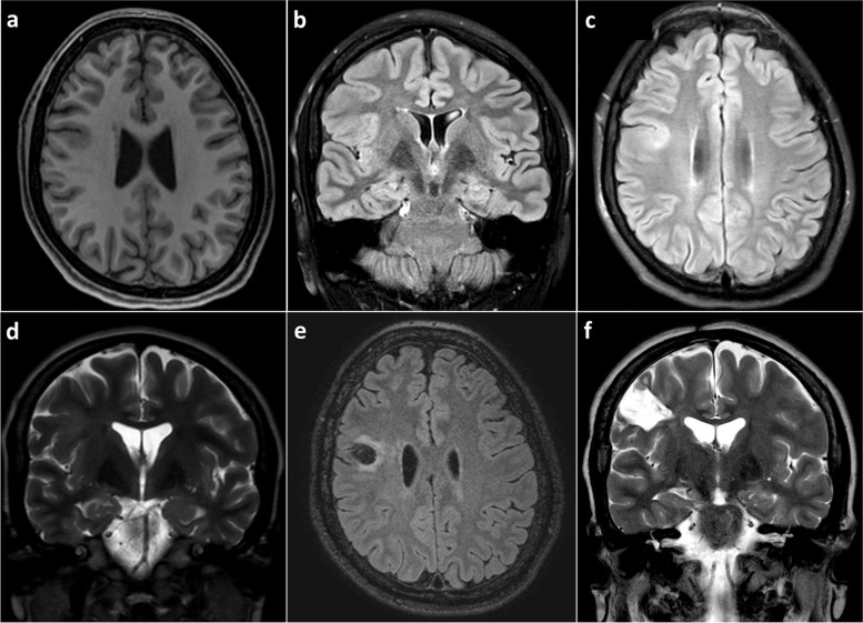
Case presentation: We report the case of a 21-year-old female with seizure onset at the age of 9, characterized by left-hand dystonic posturing and impaired awareness, which evolved to bilateral tonic-clonic seizures, evaluated in a neurological referral center in Lima, Peru. During 6-h video-EEG, interictal EEG revealing focal brief potentially ictal rhythmic discharges (BIRDs) over the right frontal central region, lasting less than 10 s. The ictal features were characterized by low-voltage fast activity over the same area. Brain magnetic resonance imaging (MRI) demonstrated a focal lesion of focal cortical dysplasia type II in the right frontal lobe. The patient underwent a lesionectomy guided by electrocorticography, which showed continuous polyspikes. BIRDs showing a brief burst of spikes lasting longer than 0.5 s, were also identified on intraoperative electrocorticography (ECoG) and helped define the extent of resection. The patient obtained an Engel Outcome Class IA at 6 years of follow-up.
Conclusions: The atypical BIRDs on ECoG can be used as a prognostic biomarker for prolonged seizure-freedom outcome in patients with epilepsy. Additional reports are needed in developing countries with and without brain MRI lesions to advance outpatient presurgical evaluations despite limited resources.
 求助内容:
求助内容: 应助结果提醒方式:


