Dua Özsoylu , Sefa Kizildag , Michael J. Schöning , Torsten Wagner
{"title":"利用等离子体功能化光寻址电位传感器(LAPS)对微流体通道内细胞外酸化进行差分化学成像","authors":"Dua Özsoylu , Sefa Kizildag , Michael J. Schöning , Torsten Wagner","doi":"10.1016/j.phmed.2020.100030","DOIUrl":null,"url":null,"abstract":"<div><p>Extracellular acidification is a basic indicator for alterations in two vital metabolic pathways: glycolysis and cellular respiration. Measuring these alterations by monitoring extracellular acidification using cell-based biosensors such as LAPS plays an important role in studying these pathways whose disorders are associated with numerous diseases including cancer. However, the surface of the biosensors must be specially tailored to ensure high cell compatibility so that cells can represent more <em>in vivo</em>-like behavior, which is critical to gain more realistic <em>in vitro</em> results from the analyses, e.g., drug discovery experiments. In this work, O<sub>2</sub> plasma patterning on the LAPS surface is studied to enhance surface features of the sensor chip, e.g., wettability and biofunctionality. The surface treated with O<sub>2</sub> plasma for 30 s exhibits enhanced cytocompatibility for adherent CHO–K1 cells, which promotes cell spreading and proliferation. The plasma-modified LAPS chip is then integrated into a microfluidic system, which provides two identical channels to facilitate differential measurements of the extracellular acidification of CHO–K1 cells. To the best of our knowledge, it is the first time that extracellular acidification within microfluidic channels is quantitatively visualized as differential (bio-)chemical images.</p></div>","PeriodicalId":37787,"journal":{"name":"Physics in Medicine","volume":"10 ","pages":"Article 100030"},"PeriodicalIF":0.0000,"publicationDate":"2020-12-01","publicationTypes":"Journal Article","fieldsOfStudy":null,"isOpenAccess":false,"openAccessPdf":"https://sci-hub-pdf.com/10.1016/j.phmed.2020.100030","citationCount":"6","resultStr":"{\"title\":\"Differential chemical imaging of extracellular acidification within microfluidic channels using a plasma-functionalized light-addressable potentiometric sensor (LAPS)\",\"authors\":\"Dua Özsoylu , Sefa Kizildag , Michael J. Schöning , Torsten Wagner\",\"doi\":\"10.1016/j.phmed.2020.100030\",\"DOIUrl\":null,\"url\":null,\"abstract\":\"<div><p>Extracellular acidification is a basic indicator for alterations in two vital metabolic pathways: glycolysis and cellular respiration. Measuring these alterations by monitoring extracellular acidification using cell-based biosensors such as LAPS plays an important role in studying these pathways whose disorders are associated with numerous diseases including cancer. However, the surface of the biosensors must be specially tailored to ensure high cell compatibility so that cells can represent more <em>in vivo</em>-like behavior, which is critical to gain more realistic <em>in vitro</em> results from the analyses, e.g., drug discovery experiments. In this work, O<sub>2</sub> plasma patterning on the LAPS surface is studied to enhance surface features of the sensor chip, e.g., wettability and biofunctionality. The surface treated with O<sub>2</sub> plasma for 30 s exhibits enhanced cytocompatibility for adherent CHO–K1 cells, which promotes cell spreading and proliferation. The plasma-modified LAPS chip is then integrated into a microfluidic system, which provides two identical channels to facilitate differential measurements of the extracellular acidification of CHO–K1 cells. To the best of our knowledge, it is the first time that extracellular acidification within microfluidic channels is quantitatively visualized as differential (bio-)chemical images.</p></div>\",\"PeriodicalId\":37787,\"journal\":{\"name\":\"Physics in Medicine\",\"volume\":\"10 \",\"pages\":\"Article 100030\"},\"PeriodicalIF\":0.0000,\"publicationDate\":\"2020-12-01\",\"publicationTypes\":\"Journal Article\",\"fieldsOfStudy\":null,\"isOpenAccess\":false,\"openAccessPdf\":\"https://sci-hub-pdf.com/10.1016/j.phmed.2020.100030\",\"citationCount\":\"6\",\"resultStr\":null,\"platform\":\"Semanticscholar\",\"paperid\":null,\"PeriodicalName\":\"Physics in Medicine\",\"FirstCategoryId\":\"1085\",\"ListUrlMain\":\"https://www.sciencedirect.com/science/article/pii/S2352451020300068\",\"RegionNum\":0,\"RegionCategory\":null,\"ArticlePicture\":[],\"TitleCN\":null,\"AbstractTextCN\":null,\"PMCID\":null,\"EPubDate\":\"\",\"PubModel\":\"\",\"JCR\":\"Q3\",\"JCRName\":\"Medicine\",\"Score\":null,\"Total\":0}","platform":"Semanticscholar","paperid":null,"PeriodicalName":"Physics in Medicine","FirstCategoryId":"1085","ListUrlMain":"https://www.sciencedirect.com/science/article/pii/S2352451020300068","RegionNum":0,"RegionCategory":null,"ArticlePicture":[],"TitleCN":null,"AbstractTextCN":null,"PMCID":null,"EPubDate":"","PubModel":"","JCR":"Q3","JCRName":"Medicine","Score":null,"Total":0}
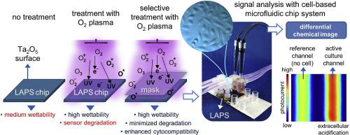
Differential chemical imaging of extracellular acidification within microfluidic channels using a plasma-functionalized light-addressable potentiometric sensor (LAPS)
Extracellular acidification is a basic indicator for alterations in two vital metabolic pathways: glycolysis and cellular respiration. Measuring these alterations by monitoring extracellular acidification using cell-based biosensors such as LAPS plays an important role in studying these pathways whose disorders are associated with numerous diseases including cancer. However, the surface of the biosensors must be specially tailored to ensure high cell compatibility so that cells can represent more in vivo-like behavior, which is critical to gain more realistic in vitro results from the analyses, e.g., drug discovery experiments. In this work, O2 plasma patterning on the LAPS surface is studied to enhance surface features of the sensor chip, e.g., wettability and biofunctionality. The surface treated with O2 plasma for 30 s exhibits enhanced cytocompatibility for adherent CHO–K1 cells, which promotes cell spreading and proliferation. The plasma-modified LAPS chip is then integrated into a microfluidic system, which provides two identical channels to facilitate differential measurements of the extracellular acidification of CHO–K1 cells. To the best of our knowledge, it is the first time that extracellular acidification within microfluidic channels is quantitatively visualized as differential (bio-)chemical images.
期刊介绍:
The scope of Physics in Medicine consists of the application of theoretical and practical physics to medicine, physiology and biology. Topics covered are: Physics of Imaging Ultrasonic imaging, Optical imaging, X-ray imaging, Fluorescence Physics of Electromagnetics Neural Engineering, Signal analysis in Medicine, Electromagnetics and the nerve system, Quantum Electronics Physics of Therapy Ultrasonic therapy, Vibrational medicine, Laser Physics Physics of Materials and Mechanics Physics of impact and injuries, Physics of proteins, Metamaterials, Nanoscience and Nanotechnology, Biomedical Materials, Physics of vascular and cerebrovascular diseases, Micromechanics and Micro engineering, Microfluidics in medicine, Mechanics of the human body, Rotary molecular motors, Biological physics, Physics of bio fabrication and regenerative medicine Physics of Instrumentation Engineering of instruments, Physical effects of the application of instruments, Measurement Science and Technology, Physics of micro-labs and bioanalytical sensor devices, Optical instrumentation, Ultrasound instruments Physics of Hearing and Seeing Acoustics and hearing, Physics of hearing aids, Optics and vision, Physics of vision aids Physics of Space Medicine Space physiology, Space medicine related Physics.

 求助内容:
求助内容: 应助结果提醒方式:


