成人肝脏未分化胚胎肉瘤的CT和MRI特征

Q4 Medicine
Jiajia Pan, Fang Chen, Hao Zhang, Q. Qiu
{"title":"成人肝脏未分化胚胎肉瘤的CT和MRI特征","authors":"Jiajia Pan, Fang Chen, Hao Zhang, Q. Qiu","doi":"10.3760/CMA.J.ISSN.1007-8118.2019.11.006","DOIUrl":null,"url":null,"abstract":"Objective \nTo analyze the CT and MRI manifestations of undifferentiated hepatic embryonal sarcoma in adults with an aim to improve the preoperative diagnosis of this disease. \n \n \nMethods \nA retrospective analysis was carried out on the clinical data of 11 patients with undifferentiated hepatic embryonal sarcoma treated in Wenzhou People’s Hospital of Zhejiang and Wenzhou Central Hospital of Zhejiang between January 2008 and December 2018. There were 5 males and 6 females. The age ranged from 46 to 76 years (median 64 years). The site, size, morphology, margin, and pattern and degree of enhancement of the tumors were analyzed. \n \n \nResults \nAmong the 11 patients, 10 patients had a single tumor, only 1 patient had multiple tumors. A total of seven patients' tumors were located in the right liver, 2 in the left liver, and 2 involved both the left and right livers. The maximum diameter of the tumors ranged from 5.5 cm to 21.2 cm, the maximum diameter (10.2±4.8 cm). The edge of tumors was clear in 9 patients and unclear in 2 patients. The tumors were elliptical in 4 patients or irregular in 7 patients. Plain CT scan showed 4 patients' tumors had mainly a cystic component, while 5 patients had both cystic and solid components. Plain MRI scan showed that five tumors were cystic and solid, with mixed internal signals. T1 weighted imaging (T1WI) showed slightly lower and lower signals; T2 weighted imaging (T2WI) showed slightly higher and higher signals. There was a low signal stripe separating inside; Diffusion-weighted imaging showed a high signal and apparent diffusion coefficient showed a low signal. The enhancement was characterized by \"fast in and fast out\" (marked enhancement in the arterial phase and fast clearance in the venous phase), and \"delayed enhancement\" (mild to moderate enhancement in the arterial phase and even more obvious enhancement in the venous or delayed phase). \n \n \nConclusion \nAdult undifferentiated embryonic sarcoma has certain CT and MRI features. Its shape is elliptical or irregular, and mostly with a clear boundary. There were intratumoral cystic or cystic solid components, with mixed density/signal on CT and MRI, being characterized by \"fast in and fast out\" and \"delayed enhancement\" on intravenous contrast enhanced scans. \n \n \nKey words: \nLiver neoplasms; Tomography, X-ray computed; Magnetic resonance imaging; Undifferentiated embryonal sarcoma","PeriodicalId":10021,"journal":{"name":"中华肝胆外科杂志","volume":"25 1","pages":"819-822"},"PeriodicalIF":0.0000,"publicationDate":"2019-11-28","publicationTypes":"Journal Article","fieldsOfStudy":null,"isOpenAccess":false,"openAccessPdf":"","citationCount":"0","resultStr":"{\"title\":\"CT and MRI features of undifferentiated embryonal sarcoma of liver in adults\",\"authors\":\"Jiajia Pan, Fang Chen, Hao Zhang, Q. Qiu\",\"doi\":\"10.3760/CMA.J.ISSN.1007-8118.2019.11.006\",\"DOIUrl\":null,\"url\":null,\"abstract\":\"Objective \\nTo analyze the CT and MRI manifestations of undifferentiated hepatic embryonal sarcoma in adults with an aim to improve the preoperative diagnosis of this disease. \\n \\n \\nMethods \\nA retrospective analysis was carried out on the clinical data of 11 patients with undifferentiated hepatic embryonal sarcoma treated in Wenzhou People’s Hospital of Zhejiang and Wenzhou Central Hospital of Zhejiang between January 2008 and December 2018. There were 5 males and 6 females. The age ranged from 46 to 76 years (median 64 years). The site, size, morphology, margin, and pattern and degree of enhancement of the tumors were analyzed. \\n \\n \\nResults \\nAmong the 11 patients, 10 patients had a single tumor, only 1 patient had multiple tumors. A total of seven patients' tumors were located in the right liver, 2 in the left liver, and 2 involved both the left and right livers. The maximum diameter of the tumors ranged from 5.5 cm to 21.2 cm, the maximum diameter (10.2±4.8 cm). The edge of tumors was clear in 9 patients and unclear in 2 patients. The tumors were elliptical in 4 patients or irregular in 7 patients. Plain CT scan showed 4 patients' tumors had mainly a cystic component, while 5 patients had both cystic and solid components. Plain MRI scan showed that five tumors were cystic and solid, with mixed internal signals. T1 weighted imaging (T1WI) showed slightly lower and lower signals; T2 weighted imaging (T2WI) showed slightly higher and higher signals. There was a low signal stripe separating inside; Diffusion-weighted imaging showed a high signal and apparent diffusion coefficient showed a low signal. The enhancement was characterized by \\\"fast in and fast out\\\" (marked enhancement in the arterial phase and fast clearance in the venous phase), and \\\"delayed enhancement\\\" (mild to moderate enhancement in the arterial phase and even more obvious enhancement in the venous or delayed phase). \\n \\n \\nConclusion \\nAdult undifferentiated embryonic sarcoma has certain CT and MRI features. Its shape is elliptical or irregular, and mostly with a clear boundary. There were intratumoral cystic or cystic solid components, with mixed density/signal on CT and MRI, being characterized by \\\"fast in and fast out\\\" and \\\"delayed enhancement\\\" on intravenous contrast enhanced scans. \\n \\n \\nKey words: \\nLiver neoplasms; Tomography, X-ray computed; Magnetic resonance imaging; Undifferentiated embryonal sarcoma\",\"PeriodicalId\":10021,\"journal\":{\"name\":\"中华肝胆外科杂志\",\"volume\":\"25 1\",\"pages\":\"819-822\"},\"PeriodicalIF\":0.0000,\"publicationDate\":\"2019-11-28\",\"publicationTypes\":\"Journal Article\",\"fieldsOfStudy\":null,\"isOpenAccess\":false,\"openAccessPdf\":\"\",\"citationCount\":\"0\",\"resultStr\":null,\"platform\":\"Semanticscholar\",\"paperid\":null,\"PeriodicalName\":\"中华肝胆外科杂志\",\"FirstCategoryId\":\"3\",\"ListUrlMain\":\"https://doi.org/10.3760/CMA.J.ISSN.1007-8118.2019.11.006\",\"RegionNum\":0,\"RegionCategory\":null,\"ArticlePicture\":[],\"TitleCN\":null,\"AbstractTextCN\":null,\"PMCID\":null,\"EPubDate\":\"\",\"PubModel\":\"\",\"JCR\":\"Q4\",\"JCRName\":\"Medicine\",\"Score\":null,\"Total\":0}","platform":"Semanticscholar","paperid":null,"PeriodicalName":"中华肝胆外科杂志","FirstCategoryId":"3","ListUrlMain":"https://doi.org/10.3760/CMA.J.ISSN.1007-8118.2019.11.006","RegionNum":0,"RegionCategory":null,"ArticlePicture":[],"TitleCN":null,"AbstractTextCN":null,"PMCID":null,"EPubDate":"","PubModel":"","JCR":"Q4","JCRName":"Medicine","Score":null,"Total":0}
引用次数: 0

摘要

目的分析成人未分化肝胚胎肉瘤的CT和MRI表现,提高对该病的术前诊断。方法回顾性分析2008年1月至2018年12月在浙江省温州市人民医院和浙江省温州市中心医院治疗的11例未分化肝胚胎肉瘤患者的临床资料。男性5人,女性6人。年龄46 ~ 76岁(中位64岁)。分析肿瘤的位置、大小、形态、边缘、类型和增强程度。结果11例患者中单发肿瘤10例,多发肿瘤1例。7例患者肿瘤位于右肝,2例位于左肝,2例同时累及左右肝。肿瘤最大直径为5.5 cm ~ 21.2 cm,最大直径(10.2±4.8 cm)。9例肿瘤边缘清晰,2例肿瘤边缘不清晰。肿瘤呈椭圆形者4例,不规则者7例。CT平扫显示4例肿瘤以囊性成分为主,5例肿瘤既有囊性成分又有实性成分。MRI平扫示5例肿瘤为囊性实性,内信号混杂。T1加权成像(T1WI)信号稍低、稍低;T2加权成像(T2WI)呈略高、高信号。里面有一条低信号条纹;弥散加权成像为高信号,视扩散系数为低信号。强化表现为“快进快出”(动脉期强化明显,静脉期清除快)和“延迟强化”(动脉期轻度至中度强化,静脉期或延迟期强化更为明显)。结论成人未分化胚胎肉瘤具有一定的CT和MRI特征。它的形状是椭圆形或不规则的,大多有一个明确的边界。肿瘤内可见囊性或囊性实性成分,CT、MRI表现为密度/信号混合,静脉增强扫描表现为“快进快出”、“延迟增强”。关键词:肝脏肿瘤;断层扫描,x射线计算机;磁共振成像;未分化胚胎肉瘤
本文章由计算机程序翻译,如有差异,请以英文原文为准。
CT and MRI features of undifferentiated embryonal sarcoma of liver in adults
Objective To analyze the CT and MRI manifestations of undifferentiated hepatic embryonal sarcoma in adults with an aim to improve the preoperative diagnosis of this disease. Methods A retrospective analysis was carried out on the clinical data of 11 patients with undifferentiated hepatic embryonal sarcoma treated in Wenzhou People’s Hospital of Zhejiang and Wenzhou Central Hospital of Zhejiang between January 2008 and December 2018. There were 5 males and 6 females. The age ranged from 46 to 76 years (median 64 years). The site, size, morphology, margin, and pattern and degree of enhancement of the tumors were analyzed. Results Among the 11 patients, 10 patients had a single tumor, only 1 patient had multiple tumors. A total of seven patients' tumors were located in the right liver, 2 in the left liver, and 2 involved both the left and right livers. The maximum diameter of the tumors ranged from 5.5 cm to 21.2 cm, the maximum diameter (10.2±4.8 cm). The edge of tumors was clear in 9 patients and unclear in 2 patients. The tumors were elliptical in 4 patients or irregular in 7 patients. Plain CT scan showed 4 patients' tumors had mainly a cystic component, while 5 patients had both cystic and solid components. Plain MRI scan showed that five tumors were cystic and solid, with mixed internal signals. T1 weighted imaging (T1WI) showed slightly lower and lower signals; T2 weighted imaging (T2WI) showed slightly higher and higher signals. There was a low signal stripe separating inside; Diffusion-weighted imaging showed a high signal and apparent diffusion coefficient showed a low signal. The enhancement was characterized by "fast in and fast out" (marked enhancement in the arterial phase and fast clearance in the venous phase), and "delayed enhancement" (mild to moderate enhancement in the arterial phase and even more obvious enhancement in the venous or delayed phase). Conclusion Adult undifferentiated embryonic sarcoma has certain CT and MRI features. Its shape is elliptical or irregular, and mostly with a clear boundary. There were intratumoral cystic or cystic solid components, with mixed density/signal on CT and MRI, being characterized by "fast in and fast out" and "delayed enhancement" on intravenous contrast enhanced scans. Key words: Liver neoplasms; Tomography, X-ray computed; Magnetic resonance imaging; Undifferentiated embryonal sarcoma
求助全文
通过发布文献求助,成功后即可免费获取论文全文。 去求助
来源期刊
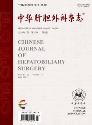
中华肝胆外科杂志
中华肝胆外科杂志 Medicine-Gastroenterology
CiteScore
0.20
自引率
0.00%
发文量
7101
期刊介绍: Chinese Journal of Hepatobiliary Surgery is an academic journal organized by the Chinese Medical Association and supervised by the China Association for Science and Technology, founded in 1995. The journal has the following columns: review, hot spotlight, academic thinking, thesis, experimental research, short thesis, case report, synthesis, etc. The journal has been recognized by Beida Journal (Chinese Journal of Humanities and Social Sciences). Chinese Journal of Hepatobiliary Surgery has been included in famous databases such as Peking University Journal (Chinese Journal of Humanities and Social Sciences), CSCD Source Journals of China Science Citation Database (with Extended Version) and so on, and it is one of the national key academic journals under the supervision of China Association for Science and Technology.
×
引用
GB/T 7714-2015
复制
MLA
复制
APA
复制
导出至
BibTeX EndNote RefMan NoteFirst NoteExpress
×
提示
您的信息不完整,为了账户安全,请先补充。
现在去补充
×
提示
您因"违规操作"
具体请查看互助需知
我知道了
×
提示
确定
请完成安全验证×
copy
已复制链接
快去分享给好友吧!
我知道了
右上角分享
点击右上角分享
0
联系我们:info@booksci.cn Book学术提供免费学术资源搜索服务,方便国内外学者检索中英文文献。致力于提供最便捷和优质的服务体验。 Copyright © 2023 布克学术 All rights reserved.
京ICP备2023020795号-1
ghs 京公网安备 11010802042870号
Book学术文献互助
Book学术文献互助群
群 号:481959085
Book学术官方微信