野生型p53诱导的磷酸酶在缺氧条件下滋养层细胞凋亡中的补偿作用

Q4 Medicine
Bin Tan, C. Tong, Yu Yuan, Li Lin
{"title":"野生型p53诱导的磷酸酶在缺氧条件下滋养层细胞凋亡中的补偿作用","authors":"Bin Tan, C. Tong, Yu Yuan, Li Lin","doi":"10.3760/CMA.J.ISSN.1007-9408.2019.10.006","DOIUrl":null,"url":null,"abstract":"Objective \nTo investigate the mechanism of wild-type p53-induced phosphatase (Wip1) in regulating p53-dependent apoptosis of trophoblasts for further understanding the etiology of preeclampsia (PE). \n \n \nMethods \nPlacenta tissues were collected from normal (n=15) and PE (n=13) gravidas who underwent caesarean section in the First Affiliated Hospital of Chongqing Medical University from June 2017 to December 2018. Chorionic villus and decidua tissues were collected from another 10 women who aborted in early pregnancy. Two in vitro trophoblastic hypoxia cultures were established by subjecting human chorionic trophoblast cells (HTR8/SVneo) to either hypoxia intervention in incubator (HII) or simulated ischemic buffer (SIB). Wip1 expressions at the transcriptional and protein levels were determined by real-time quantitative polymerase chain reaction and Western blotting, respectively. The localization of Wip1 in placental tissues and HTR8/SVneo cells was determined by immunohistochemistry and immunofluorescence. Cell apoptosis was assessed by flow cytometry after viral infection and hypoxia. And the changes of pathway-related molecules including p53, phospho-p53 (p-p53), mouse double minute 2 homolog (Mdm2) and cleaved caspase3 (cl-cas3) were measured by Western blotting. The impact of Wip1 on Mdm2-p53 interaction was examined by co-immunoprecipitation. NVP-CGM097, an Mdm2-p53 specific inhibitor, was administered in PE cell models to verify the regulation of Wip1 on trophoblastic apoptosis through Mdm2-p53 pathway. Independent student's t-test, Welch's t-test and one-way analysis of variance were used as statistical methods. \n \n \nResults \n(1) Wip1 expression, which was mainly in trophoblast cells, was significantly elevated in human PE placentas (mRNA: 1.711±0.141 vs 0.860±0.126, t=4.496; protein: 0.449±0.027 vs 0.192±0.019, t=7.902) and in both in vitro trophoblastic PE models (protein in HII: 1.376±0.086 vs 0.977±0.114, t=2.792; SIB: 1.243±0.057 vs 0.381±0.045, t=11.910) compared with the corresponding control groups (all P<0.05). (2) Compared with corresponding control groups, overexpression of Wip1 suppressed the hypoxia-induced upregulation of p53 (HII: 0.185±0.024 vs 0.572±0.072; SIB: 0.400±0.067 vs 0.803±0.064), cl-cas3 (HII: 0.243±0.034 vs 0.529±0.072; SIB: 0.179±0.011 vs 0.368±0.025) and p-p53/p53 protein expression (HII: 1.326±0.129 vs 2.100±0.187; SIB: 0.473±0.028 vs 0.925±0.036) and also reduced the apoptosis rate [HII: (8.925±1.092)% vs (17.610±1.980)%; SIB: (13.910±1.886)% vs (24.650±1.622)%], which in turn promoted Mdm2-p53 binding (all P<0.05). However, knockdown of Wip1 gene expression in HTR8/SVneo cells brought about opposite effects (all P<0.05). (3) Neither overexpression nor knockdown of Wip1 influenced p53 or cl-cas3 expression when Mdm2-p53 interaction was blocked by NVP-CGM097. \n \n \nConclusions \nMdm2-p53 interaction promoted by Wip1 upregulation could compensate for the trophoblastic p53 accumulation in response to hypoxia, while exogenous upregulation of Wip1 in trophoblasts may reverse hypoxia-induced apoptosis. Therefore, this might provide a new therapeutic target for PE. \n \n \nKey words: \nPre-eclampsia; Cell hypoxia; Trophoblasts; Protein phosphatase 2C; Apoptosis","PeriodicalId":52320,"journal":{"name":"中华围产医学杂志","volume":"22 1","pages":"712-721"},"PeriodicalIF":0.0000,"publicationDate":"2019-10-16","publicationTypes":"Journal Article","fieldsOfStudy":null,"isOpenAccess":false,"openAccessPdf":"","citationCount":"0","resultStr":"{\"title\":\"Compensatory role of wild-type p53-induced phosphatase in trophoblastic apoptosis in response to hypoxia\",\"authors\":\"Bin Tan, C. Tong, Yu Yuan, Li Lin\",\"doi\":\"10.3760/CMA.J.ISSN.1007-9408.2019.10.006\",\"DOIUrl\":null,\"url\":null,\"abstract\":\"Objective \\nTo investigate the mechanism of wild-type p53-induced phosphatase (Wip1) in regulating p53-dependent apoptosis of trophoblasts for further understanding the etiology of preeclampsia (PE). \\n \\n \\nMethods \\nPlacenta tissues were collected from normal (n=15) and PE (n=13) gravidas who underwent caesarean section in the First Affiliated Hospital of Chongqing Medical University from June 2017 to December 2018. Chorionic villus and decidua tissues were collected from another 10 women who aborted in early pregnancy. Two in vitro trophoblastic hypoxia cultures were established by subjecting human chorionic trophoblast cells (HTR8/SVneo) to either hypoxia intervention in incubator (HII) or simulated ischemic buffer (SIB). Wip1 expressions at the transcriptional and protein levels were determined by real-time quantitative polymerase chain reaction and Western blotting, respectively. The localization of Wip1 in placental tissues and HTR8/SVneo cells was determined by immunohistochemistry and immunofluorescence. Cell apoptosis was assessed by flow cytometry after viral infection and hypoxia. And the changes of pathway-related molecules including p53, phospho-p53 (p-p53), mouse double minute 2 homolog (Mdm2) and cleaved caspase3 (cl-cas3) were measured by Western blotting. The impact of Wip1 on Mdm2-p53 interaction was examined by co-immunoprecipitation. NVP-CGM097, an Mdm2-p53 specific inhibitor, was administered in PE cell models to verify the regulation of Wip1 on trophoblastic apoptosis through Mdm2-p53 pathway. Independent student's t-test, Welch's t-test and one-way analysis of variance were used as statistical methods. \\n \\n \\nResults \\n(1) Wip1 expression, which was mainly in trophoblast cells, was significantly elevated in human PE placentas (mRNA: 1.711±0.141 vs 0.860±0.126, t=4.496; protein: 0.449±0.027 vs 0.192±0.019, t=7.902) and in both in vitro trophoblastic PE models (protein in HII: 1.376±0.086 vs 0.977±0.114, t=2.792; SIB: 1.243±0.057 vs 0.381±0.045, t=11.910) compared with the corresponding control groups (all P<0.05). (2) Compared with corresponding control groups, overexpression of Wip1 suppressed the hypoxia-induced upregulation of p53 (HII: 0.185±0.024 vs 0.572±0.072; SIB: 0.400±0.067 vs 0.803±0.064), cl-cas3 (HII: 0.243±0.034 vs 0.529±0.072; SIB: 0.179±0.011 vs 0.368±0.025) and p-p53/p53 protein expression (HII: 1.326±0.129 vs 2.100±0.187; SIB: 0.473±0.028 vs 0.925±0.036) and also reduced the apoptosis rate [HII: (8.925±1.092)% vs (17.610±1.980)%; SIB: (13.910±1.886)% vs (24.650±1.622)%], which in turn promoted Mdm2-p53 binding (all P<0.05). However, knockdown of Wip1 gene expression in HTR8/SVneo cells brought about opposite effects (all P<0.05). (3) Neither overexpression nor knockdown of Wip1 influenced p53 or cl-cas3 expression when Mdm2-p53 interaction was blocked by NVP-CGM097. \\n \\n \\nConclusions \\nMdm2-p53 interaction promoted by Wip1 upregulation could compensate for the trophoblastic p53 accumulation in response to hypoxia, while exogenous upregulation of Wip1 in trophoblasts may reverse hypoxia-induced apoptosis. Therefore, this might provide a new therapeutic target for PE. \\n \\n \\nKey words: \\nPre-eclampsia; Cell hypoxia; Trophoblasts; Protein phosphatase 2C; Apoptosis\",\"PeriodicalId\":52320,\"journal\":{\"name\":\"中华围产医学杂志\",\"volume\":\"22 1\",\"pages\":\"712-721\"},\"PeriodicalIF\":0.0000,\"publicationDate\":\"2019-10-16\",\"publicationTypes\":\"Journal Article\",\"fieldsOfStudy\":null,\"isOpenAccess\":false,\"openAccessPdf\":\"\",\"citationCount\":\"0\",\"resultStr\":null,\"platform\":\"Semanticscholar\",\"paperid\":null,\"PeriodicalName\":\"中华围产医学杂志\",\"FirstCategoryId\":\"3\",\"ListUrlMain\":\"https://doi.org/10.3760/CMA.J.ISSN.1007-9408.2019.10.006\",\"RegionNum\":0,\"RegionCategory\":null,\"ArticlePicture\":[],\"TitleCN\":null,\"AbstractTextCN\":null,\"PMCID\":null,\"EPubDate\":\"\",\"PubModel\":\"\",\"JCR\":\"Q4\",\"JCRName\":\"Medicine\",\"Score\":null,\"Total\":0}","platform":"Semanticscholar","paperid":null,"PeriodicalName":"中华围产医学杂志","FirstCategoryId":"3","ListUrlMain":"https://doi.org/10.3760/CMA.J.ISSN.1007-9408.2019.10.006","RegionNum":0,"RegionCategory":null,"ArticlePicture":[],"TitleCN":null,"AbstractTextCN":null,"PMCID":null,"EPubDate":"","PubModel":"","JCR":"Q4","JCRName":"Medicine","Score":null,"Total":0}
引用次数: 0

摘要

目的探讨野生型p53诱导的磷酸酶(Wip1)调节p53依赖性滋养层细胞凋亡的机制,以进一步了解子痫前期(PE)的病因。方法收集2017年6月至2018年12月在重庆医科大学第一附属医院剖腹产的正常(n=15)和PE(n=13)孕妇的胎盘组织。从另外10名早孕流产妇女身上采集绒毛和蜕膜组织。通过在培养箱中对人绒毛膜滋养层细胞(HTR8/SVneo)进行缺氧干预(HII)或模拟缺血缓冲液(SIB),建立了两种体外滋养层缺氧培养物。分别通过实时定量聚合酶链反应和蛋白质印迹测定转录和蛋白质水平上的Wip1表达。通过免疫组织化学和免疫荧光测定Wip1在胎盘组织和HTR8/SVneo细胞中的定位。通过流式细胞术评估病毒感染和缺氧后的细胞凋亡。Western印迹法检测p53、磷酸化p53(p-p53)、小鼠双分钟2同源物(Mdm2)和裂解caspase3(cl-cas3)等通路相关分子的变化。通过共免疫沉淀检测Wip1对Mdm2-p53相互作用的影响。NVP-CGM097,一种Mdm2-p53特异性抑制剂,在PE细胞模型中给药,以验证Wip1通过Mdm2-p53途径对滋养层细胞凋亡的调节。采用独立学生t检验、韦尔奇t检验和单因素方差分析作为统计方法。结果(1)Wip1主要在滋养层细胞中表达,与相应的对照组相比,在人PE胎盘中显著升高(mRNA:1.711±0.141 vs 0.860±0.126,t=4.496;蛋白质:0.449±0.027 vs 0.192±0.019,t=7.902),在两种体外滋养层PE模型中(HII:1.376±0.086 vs 0.977±0.114,t=2.792;SIB:1.243±0.057 vs 0.381±0.045,t=11.910)(均P<0.05),Wip1的过表达抑制了缺氧诱导的p53的上调(HII:0.185±0.024 vs 0.572±0.072;SIB:0.400±0.067 vs 0.803±0.064),cl-cas3(HII:0.243±0.034 vs 0.529±0.072;SIB:0.179±0.011 vs 0.368±0.025)和p-p53/p53蛋白表达(HII:1.326±0.129 vs 2.100±0.187;SIB:0.473±0.028 vs 0.925±0.036),还降低了细胞凋亡率[HII:(8.925±1.092)%vs(17.610±1.980)%;SIB:(13.910±1.886)%vs,(3)NVP-CGM097阻断Mdm2-p53相互作用时,Wip1的过表达和敲低均不影响p53和cl-cas3的表达。结论Wip1上调所促进的Mdm2-p53相互作用可以补偿滋养层细胞缺氧时p53的积累,而Wip1在滋养层中的外源性上调可能逆转缺氧诱导的细胞凋亡。因此,这可能为PE提供一个新的治疗靶点;细胞缺氧;滋养层;蛋白磷酸酶2C;细胞凋亡
本文章由计算机程序翻译,如有差异,请以英文原文为准。
Compensatory role of wild-type p53-induced phosphatase in trophoblastic apoptosis in response to hypoxia
Objective To investigate the mechanism of wild-type p53-induced phosphatase (Wip1) in regulating p53-dependent apoptosis of trophoblasts for further understanding the etiology of preeclampsia (PE). Methods Placenta tissues were collected from normal (n=15) and PE (n=13) gravidas who underwent caesarean section in the First Affiliated Hospital of Chongqing Medical University from June 2017 to December 2018. Chorionic villus and decidua tissues were collected from another 10 women who aborted in early pregnancy. Two in vitro trophoblastic hypoxia cultures were established by subjecting human chorionic trophoblast cells (HTR8/SVneo) to either hypoxia intervention in incubator (HII) or simulated ischemic buffer (SIB). Wip1 expressions at the transcriptional and protein levels were determined by real-time quantitative polymerase chain reaction and Western blotting, respectively. The localization of Wip1 in placental tissues and HTR8/SVneo cells was determined by immunohistochemistry and immunofluorescence. Cell apoptosis was assessed by flow cytometry after viral infection and hypoxia. And the changes of pathway-related molecules including p53, phospho-p53 (p-p53), mouse double minute 2 homolog (Mdm2) and cleaved caspase3 (cl-cas3) were measured by Western blotting. The impact of Wip1 on Mdm2-p53 interaction was examined by co-immunoprecipitation. NVP-CGM097, an Mdm2-p53 specific inhibitor, was administered in PE cell models to verify the regulation of Wip1 on trophoblastic apoptosis through Mdm2-p53 pathway. Independent student's t-test, Welch's t-test and one-way analysis of variance were used as statistical methods. Results (1) Wip1 expression, which was mainly in trophoblast cells, was significantly elevated in human PE placentas (mRNA: 1.711±0.141 vs 0.860±0.126, t=4.496; protein: 0.449±0.027 vs 0.192±0.019, t=7.902) and in both in vitro trophoblastic PE models (protein in HII: 1.376±0.086 vs 0.977±0.114, t=2.792; SIB: 1.243±0.057 vs 0.381±0.045, t=11.910) compared with the corresponding control groups (all P<0.05). (2) Compared with corresponding control groups, overexpression of Wip1 suppressed the hypoxia-induced upregulation of p53 (HII: 0.185±0.024 vs 0.572±0.072; SIB: 0.400±0.067 vs 0.803±0.064), cl-cas3 (HII: 0.243±0.034 vs 0.529±0.072; SIB: 0.179±0.011 vs 0.368±0.025) and p-p53/p53 protein expression (HII: 1.326±0.129 vs 2.100±0.187; SIB: 0.473±0.028 vs 0.925±0.036) and also reduced the apoptosis rate [HII: (8.925±1.092)% vs (17.610±1.980)%; SIB: (13.910±1.886)% vs (24.650±1.622)%], which in turn promoted Mdm2-p53 binding (all P<0.05). However, knockdown of Wip1 gene expression in HTR8/SVneo cells brought about opposite effects (all P<0.05). (3) Neither overexpression nor knockdown of Wip1 influenced p53 or cl-cas3 expression when Mdm2-p53 interaction was blocked by NVP-CGM097. Conclusions Mdm2-p53 interaction promoted by Wip1 upregulation could compensate for the trophoblastic p53 accumulation in response to hypoxia, while exogenous upregulation of Wip1 in trophoblasts may reverse hypoxia-induced apoptosis. Therefore, this might provide a new therapeutic target for PE. Key words: Pre-eclampsia; Cell hypoxia; Trophoblasts; Protein phosphatase 2C; Apoptosis
求助全文
通过发布文献求助,成功后即可免费获取论文全文。 去求助
来源期刊
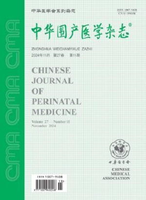
中华围产医学杂志
中华围产医学杂志 Medicine-Obstetrics and Gynecology
CiteScore
0.70
自引率
0.00%
发文量
4446
期刊介绍:
×
引用
GB/T 7714-2015
复制
MLA
复制
APA
复制
导出至
BibTeX EndNote RefMan NoteFirst NoteExpress
×
提示
您的信息不完整,为了账户安全,请先补充。
现在去补充
×
提示
您因"违规操作"
具体请查看互助需知
我知道了
×
提示
确定
请完成安全验证×
copy
已复制链接
快去分享给好友吧!
我知道了
右上角分享
点击右上角分享
0
联系我们:info@booksci.cn Book学术提供免费学术资源搜索服务,方便国内外学者检索中英文文献。致力于提供最便捷和优质的服务体验。 Copyright © 2023 布克学术 All rights reserved.
京ICP备2023020795号-1
ghs 京公网安备 11010802042870号
Book学术文献互助
Book学术文献互助群
群 号:481959085
Book学术官方微信