Jason J Chang, Casey Chen, Joe Chang, Sreenivas Koka, Jesse V Jokerst
{"title":"龈下牙石成像工具的叙述性综述。","authors":"Jason J Chang, Casey Chen, Joe Chang, Sreenivas Koka, Jesse V Jokerst","doi":"10.21037/fomm-21-57","DOIUrl":null,"url":null,"abstract":"<p><strong>Background and objective: </strong>The conventional method of detecting subgingival calculus involves using a periodontal probe to sense tactile differences on the dental root surface. Although efficient, this method can result in false positives and false negatives. This literature review explores alternative detection techniques that can detect subgingival calculus with improved accuracy and consistency. The accumulation of dental calculus below the gingival margin can foster periodontitis-inducing bacterial growth. Conventional methods of locating subgingival calculus are often inaccurate and highly dependent on clinician skill. This literature review evaluates techniques used to improve the accuracy of imaging and detecting subgingival calculus.</p><p><strong>Methods: </strong>Google Scholar, PubMed and PubMed Central databases were searched for peer-reviewed original articles evaluating subgingival calculus imaging and detection techniques. A total of 46 relevant articles ranging from 1981 to 2021 were included.</p><p><strong>Key content and findings: </strong>This narrative review discusses the subgingival calculus detection and imaging capabilities of periodontal endoscopy in an <i>in vivo</i> study and of optical coherence tomography (OCT), fluorescence spectroscopy, and differential reflectometry in <i>in vitro</i> settings. Each technique has unique benefits and limitations that distinguishes it from the others.</p><p><strong>Conclusions: </strong><i>In vitro</i> studies have revealed that techniques including periodontal endoscopy, OCT, fluorescence spectroscopy, or differential reflectometry allow for a more accurate diagnosis of subgingival calculus deposits in comparison to detection via periodontal probing. Despite the improved results, the common limitations of these techniques include longer operation times and expensive equipment. Further studies are needed to transition these imaging and detection methods to clinical environments.</p>","PeriodicalId":93098,"journal":{"name":"Frontiers of oral and maxillofacial medicine","volume":"5 ","pages":""},"PeriodicalIF":0.0000,"publicationDate":"2023-03-10","publicationTypes":"Journal Article","fieldsOfStudy":null,"isOpenAccess":false,"openAccessPdf":"https://ftp.ncbi.nlm.nih.gov/pub/pmc/oa_pdf/21/8e/nihms-1884528.PMC10569434.pdf","citationCount":"1","resultStr":"{\"title\":\"A narrative review of imaging tools for imaging subgingival calculus.\",\"authors\":\"Jason J Chang, Casey Chen, Joe Chang, Sreenivas Koka, Jesse V Jokerst\",\"doi\":\"10.21037/fomm-21-57\",\"DOIUrl\":null,\"url\":null,\"abstract\":\"<p><strong>Background and objective: </strong>The conventional method of detecting subgingival calculus involves using a periodontal probe to sense tactile differences on the dental root surface. Although efficient, this method can result in false positives and false negatives. This literature review explores alternative detection techniques that can detect subgingival calculus with improved accuracy and consistency. The accumulation of dental calculus below the gingival margin can foster periodontitis-inducing bacterial growth. Conventional methods of locating subgingival calculus are often inaccurate and highly dependent on clinician skill. This literature review evaluates techniques used to improve the accuracy of imaging and detecting subgingival calculus.</p><p><strong>Methods: </strong>Google Scholar, PubMed and PubMed Central databases were searched for peer-reviewed original articles evaluating subgingival calculus imaging and detection techniques. A total of 46 relevant articles ranging from 1981 to 2021 were included.</p><p><strong>Key content and findings: </strong>This narrative review discusses the subgingival calculus detection and imaging capabilities of periodontal endoscopy in an <i>in vivo</i> study and of optical coherence tomography (OCT), fluorescence spectroscopy, and differential reflectometry in <i>in vitro</i> settings. Each technique has unique benefits and limitations that distinguishes it from the others.</p><p><strong>Conclusions: </strong><i>In vitro</i> studies have revealed that techniques including periodontal endoscopy, OCT, fluorescence spectroscopy, or differential reflectometry allow for a more accurate diagnosis of subgingival calculus deposits in comparison to detection via periodontal probing. Despite the improved results, the common limitations of these techniques include longer operation times and expensive equipment. Further studies are needed to transition these imaging and detection methods to clinical environments.</p>\",\"PeriodicalId\":93098,\"journal\":{\"name\":\"Frontiers of oral and maxillofacial medicine\",\"volume\":\"5 \",\"pages\":\"\"},\"PeriodicalIF\":0.0000,\"publicationDate\":\"2023-03-10\",\"publicationTypes\":\"Journal Article\",\"fieldsOfStudy\":null,\"isOpenAccess\":false,\"openAccessPdf\":\"https://ftp.ncbi.nlm.nih.gov/pub/pmc/oa_pdf/21/8e/nihms-1884528.PMC10569434.pdf\",\"citationCount\":\"1\",\"resultStr\":null,\"platform\":\"Semanticscholar\",\"paperid\":null,\"PeriodicalName\":\"Frontiers of oral and maxillofacial medicine\",\"FirstCategoryId\":\"1085\",\"ListUrlMain\":\"https://doi.org/10.21037/fomm-21-57\",\"RegionNum\":0,\"RegionCategory\":null,\"ArticlePicture\":[],\"TitleCN\":null,\"AbstractTextCN\":null,\"PMCID\":null,\"EPubDate\":\"2021/10/21 0:00:00\",\"PubModel\":\"Epub\",\"JCR\":\"\",\"JCRName\":\"\",\"Score\":null,\"Total\":0}","platform":"Semanticscholar","paperid":null,"PeriodicalName":"Frontiers of oral and maxillofacial medicine","FirstCategoryId":"1085","ListUrlMain":"https://doi.org/10.21037/fomm-21-57","RegionNum":0,"RegionCategory":null,"ArticlePicture":[],"TitleCN":null,"AbstractTextCN":null,"PMCID":null,"EPubDate":"2021/10/21 0:00:00","PubModel":"Epub","JCR":"","JCRName":"","Score":null,"Total":0}
A narrative review of imaging tools for imaging subgingival calculus.
Background and objective: The conventional method of detecting subgingival calculus involves using a periodontal probe to sense tactile differences on the dental root surface. Although efficient, this method can result in false positives and false negatives. This literature review explores alternative detection techniques that can detect subgingival calculus with improved accuracy and consistency. The accumulation of dental calculus below the gingival margin can foster periodontitis-inducing bacterial growth. Conventional methods of locating subgingival calculus are often inaccurate and highly dependent on clinician skill. This literature review evaluates techniques used to improve the accuracy of imaging and detecting subgingival calculus.
Methods: Google Scholar, PubMed and PubMed Central databases were searched for peer-reviewed original articles evaluating subgingival calculus imaging and detection techniques. A total of 46 relevant articles ranging from 1981 to 2021 were included.
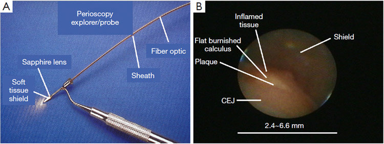
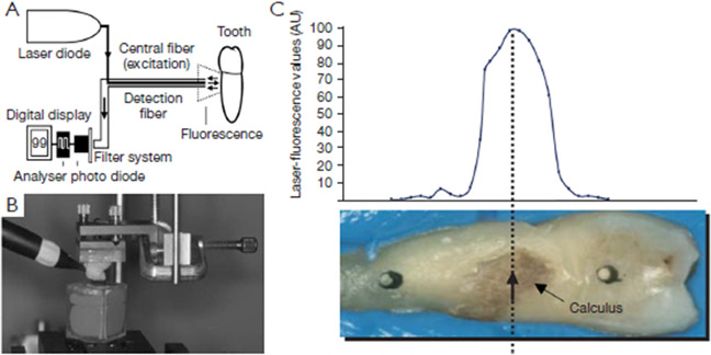
Key content and findings: This narrative review discusses the subgingival calculus detection and imaging capabilities of periodontal endoscopy in an in vivo study and of optical coherence tomography (OCT), fluorescence spectroscopy, and differential reflectometry in in vitro settings. Each technique has unique benefits and limitations that distinguishes it from the others.
Conclusions: In vitro studies have revealed that techniques including periodontal endoscopy, OCT, fluorescence spectroscopy, or differential reflectometry allow for a more accurate diagnosis of subgingival calculus deposits in comparison to detection via periodontal probing. Despite the improved results, the common limitations of these techniques include longer operation times and expensive equipment. Further studies are needed to transition these imaging and detection methods to clinical environments.


 求助内容:
求助内容: 应助结果提醒方式:


