{"title":"通过手工制作和深度学习特征相结合来识别肝转移的原发性肿瘤起源部位。","authors":"Chuheng Chen, Cheng Lu, Vidya Viswanathan, Brandon Maveal, Bhunesh Maheshwari, Joseph Willis, Anant Madabhushi","doi":"10.1002/cjp2.344","DOIUrl":null,"url":null,"abstract":"<p>Liver is one of the most common sites for metastases, which can occur on account of primary tumors from multiple sites of origin. Identifying the primary site of origin (PSO) of a metastasis can help in guiding therapeutic options for liver metastases. In this pilot study, we hypothesized that computer extracted handcrafted (HC) histomorphometric features can be utilized to identify the PSO of liver metastases. Cellular features, including tumor nuclei morphological and graph features as well as cytoplasm texture features, were extracted by computer algorithms from 175 slides (114 patients). The study comprised three experiments: (1) comparing and (2) fusing a machine learning (ML) model trained with HC pathomic features and deep learning (DL)-based classifiers to predict site of origin; (3) identifying the section of the primary tumor from which metastases were derived. For experiment 1, we divided the cohort into training sets composed of primary and matched liver metastases [60 patients, 121 whole slide images (WSIs)], and a hold-out validation set (54 patients, 54 WSIs) composed solely of liver metastases of known site of origin. Using the extracted HC features of the training set, a combination of supervised machine classifiers and unsupervised clustering was applied to identify the PSO. A random forest classifier achieved areas under the curve (AUCs) of 0.83, 0.64, 0.82, and 0.64 in classifying the metastatic tumor from colon, esophagus, breast, and pancreas on the validation set. The top features related to nuclear and peri-nuclear shape and textural attributes. We also trained a DL network to serve as a direct comparison to our method. The DL model achieved AUCs for colon: 0.94, esophagus: 0.66, breast: 0.79, and pancreas: 0.67 in identifying PSO. A decision fusion-based strategy was deployed to fuse the trained ML and DL classifiers and achieved slightly better results than ML or DL classifier alone (colon: 0.93, esophagus: 0.68, breast: 0.81, and pancreas: 0.69). For the third experiment, WSI-level attention maps were also generated using a trained DL network to generate a composite feature similarity heat map between paired primaries and their associated metastases. Our experiments revealed that epithelium-rich and moderately differentiated tumor regions of primary tumors were quantitatively similar to paired metastatic tumors. Our findings suggest that a combination of HC and DL features could potentially help identify the PSO for liver metastases while at the same time also potentially identify the spatial sites of origin for the metastases within primary tumors.</p>","PeriodicalId":48612,"journal":{"name":"Journal of Pathology Clinical Research","volume":"10 1","pages":""},"PeriodicalIF":3.7000,"publicationDate":"2023-10-11","publicationTypes":"Journal Article","fieldsOfStudy":null,"isOpenAccess":false,"openAccessPdf":"https://onlinelibrary.wiley.com/doi/epdf/10.1002/cjp2.344","citationCount":"0","resultStr":"{\"title\":\"Identifying primary tumor site of origin for liver metastases via a combination of handcrafted and deep learning features\",\"authors\":\"Chuheng Chen, Cheng Lu, Vidya Viswanathan, Brandon Maveal, Bhunesh Maheshwari, Joseph Willis, Anant Madabhushi\",\"doi\":\"10.1002/cjp2.344\",\"DOIUrl\":null,\"url\":null,\"abstract\":\"<p>Liver is one of the most common sites for metastases, which can occur on account of primary tumors from multiple sites of origin. Identifying the primary site of origin (PSO) of a metastasis can help in guiding therapeutic options for liver metastases. In this pilot study, we hypothesized that computer extracted handcrafted (HC) histomorphometric features can be utilized to identify the PSO of liver metastases. Cellular features, including tumor nuclei morphological and graph features as well as cytoplasm texture features, were extracted by computer algorithms from 175 slides (114 patients). The study comprised three experiments: (1) comparing and (2) fusing a machine learning (ML) model trained with HC pathomic features and deep learning (DL)-based classifiers to predict site of origin; (3) identifying the section of the primary tumor from which metastases were derived. For experiment 1, we divided the cohort into training sets composed of primary and matched liver metastases [60 patients, 121 whole slide images (WSIs)], and a hold-out validation set (54 patients, 54 WSIs) composed solely of liver metastases of known site of origin. Using the extracted HC features of the training set, a combination of supervised machine classifiers and unsupervised clustering was applied to identify the PSO. A random forest classifier achieved areas under the curve (AUCs) of 0.83, 0.64, 0.82, and 0.64 in classifying the metastatic tumor from colon, esophagus, breast, and pancreas on the validation set. The top features related to nuclear and peri-nuclear shape and textural attributes. We also trained a DL network to serve as a direct comparison to our method. The DL model achieved AUCs for colon: 0.94, esophagus: 0.66, breast: 0.79, and pancreas: 0.67 in identifying PSO. A decision fusion-based strategy was deployed to fuse the trained ML and DL classifiers and achieved slightly better results than ML or DL classifier alone (colon: 0.93, esophagus: 0.68, breast: 0.81, and pancreas: 0.69). For the third experiment, WSI-level attention maps were also generated using a trained DL network to generate a composite feature similarity heat map between paired primaries and their associated metastases. Our experiments revealed that epithelium-rich and moderately differentiated tumor regions of primary tumors were quantitatively similar to paired metastatic tumors. Our findings suggest that a combination of HC and DL features could potentially help identify the PSO for liver metastases while at the same time also potentially identify the spatial sites of origin for the metastases within primary tumors.</p>\",\"PeriodicalId\":48612,\"journal\":{\"name\":\"Journal of Pathology Clinical Research\",\"volume\":\"10 1\",\"pages\":\"\"},\"PeriodicalIF\":3.7000,\"publicationDate\":\"2023-10-11\",\"publicationTypes\":\"Journal Article\",\"fieldsOfStudy\":null,\"isOpenAccess\":false,\"openAccessPdf\":\"https://onlinelibrary.wiley.com/doi/epdf/10.1002/cjp2.344\",\"citationCount\":\"0\",\"resultStr\":null,\"platform\":\"Semanticscholar\",\"paperid\":null,\"PeriodicalName\":\"Journal of Pathology Clinical Research\",\"FirstCategoryId\":\"3\",\"ListUrlMain\":\"https://onlinelibrary.wiley.com/doi/10.1002/cjp2.344\",\"RegionNum\":2,\"RegionCategory\":\"医学\",\"ArticlePicture\":[],\"TitleCN\":null,\"AbstractTextCN\":null,\"PMCID\":null,\"EPubDate\":\"\",\"PubModel\":\"\",\"JCR\":\"Q1\",\"JCRName\":\"PATHOLOGY\",\"Score\":null,\"Total\":0}","platform":"Semanticscholar","paperid":null,"PeriodicalName":"Journal of Pathology Clinical Research","FirstCategoryId":"3","ListUrlMain":"https://onlinelibrary.wiley.com/doi/10.1002/cjp2.344","RegionNum":2,"RegionCategory":"医学","ArticlePicture":[],"TitleCN":null,"AbstractTextCN":null,"PMCID":null,"EPubDate":"","PubModel":"","JCR":"Q1","JCRName":"PATHOLOGY","Score":null,"Total":0}
Identifying primary tumor site of origin for liver metastases via a combination of handcrafted and deep learning features
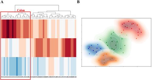
Liver is one of the most common sites for metastases, which can occur on account of primary tumors from multiple sites of origin. Identifying the primary site of origin (PSO) of a metastasis can help in guiding therapeutic options for liver metastases. In this pilot study, we hypothesized that computer extracted handcrafted (HC) histomorphometric features can be utilized to identify the PSO of liver metastases. Cellular features, including tumor nuclei morphological and graph features as well as cytoplasm texture features, were extracted by computer algorithms from 175 slides (114 patients). The study comprised three experiments: (1) comparing and (2) fusing a machine learning (ML) model trained with HC pathomic features and deep learning (DL)-based classifiers to predict site of origin; (3) identifying the section of the primary tumor from which metastases were derived. For experiment 1, we divided the cohort into training sets composed of primary and matched liver metastases [60 patients, 121 whole slide images (WSIs)], and a hold-out validation set (54 patients, 54 WSIs) composed solely of liver metastases of known site of origin. Using the extracted HC features of the training set, a combination of supervised machine classifiers and unsupervised clustering was applied to identify the PSO. A random forest classifier achieved areas under the curve (AUCs) of 0.83, 0.64, 0.82, and 0.64 in classifying the metastatic tumor from colon, esophagus, breast, and pancreas on the validation set. The top features related to nuclear and peri-nuclear shape and textural attributes. We also trained a DL network to serve as a direct comparison to our method. The DL model achieved AUCs for colon: 0.94, esophagus: 0.66, breast: 0.79, and pancreas: 0.67 in identifying PSO. A decision fusion-based strategy was deployed to fuse the trained ML and DL classifiers and achieved slightly better results than ML or DL classifier alone (colon: 0.93, esophagus: 0.68, breast: 0.81, and pancreas: 0.69). For the third experiment, WSI-level attention maps were also generated using a trained DL network to generate a composite feature similarity heat map between paired primaries and their associated metastases. Our experiments revealed that epithelium-rich and moderately differentiated tumor regions of primary tumors were quantitatively similar to paired metastatic tumors. Our findings suggest that a combination of HC and DL features could potentially help identify the PSO for liver metastases while at the same time also potentially identify the spatial sites of origin for the metastases within primary tumors.
期刊介绍:
The Journal of Pathology: Clinical Research and The Journal of Pathology serve as translational bridges between basic biomedical science and clinical medicine with particular emphasis on, but not restricted to, tissue based studies.
The focus of The Journal of Pathology: Clinical Research is the publication of studies that illuminate the clinical relevance of research in the broad area of the study of disease. Appropriately powered and validated studies with novel diagnostic, prognostic and predictive significance, and biomarker discover and validation, will be welcomed. Studies with a predominantly mechanistic basis will be more appropriate for the companion Journal of Pathology.
 求助内容:
求助内容: 应助结果提醒方式:


