{"title":"肺半定量测量和纵隔脂肪组织体积能否预测特发性肺纤维化(IPF)患者的预后?基于CT的初步研究。","authors":"Hüseyin Akkaya, Özlem Erçen Diken","doi":"10.5578/tt.20239702","DOIUrl":null,"url":null,"abstract":"<p><strong>Introduction: </strong>The aim of this study was to assess the potential of subcutaneous adipose tissue volume, mediastinal adipose tissue volume, lung density, and lung volume (as measured on high-resolution computed tomography) to predict disease progression in patients with idiopathic pulmonary fibrosis (IPF). Additionally, the study aimed to evaluate the changes in these semiquantitative measures over time.</p><p><strong>Materials and methods: </strong>The HRCT images of 57 patients diagnosed with IPF were retrospectively screened. Subcutaneous adipose tissue volume, mediastinal adipose tissue volume, and mean lung density and volume were measured at the time of diagnosis and at the 12th month. The ability of these parameters to predict progression was evaluated using the univariate and multivariate Cox regression analyses.</p><p><strong>Results: </strong>Low mediastinal adipose tissue volume at diagnosis had a 0.991-fold effect [odds ratio (OR)= 0.991, 95% confidence interval (CI)= 0.984-0.997, p< 0.001] on progression. Low mediastinal adipose tissue volume at diagnosis had a 0.993-fold effect [odds ratio (OR)= 0.993, 95% confidence interval (CI)= 0.975-1.011, p< 0.001] and progression development at the 12th month had a 6.5-fold effect [odds ratio (OR)= 6.516, 95% confidence interval (CI)= 1.594-26.639, p< 0.009] on mortality.</p><p><strong>Conclusion: </strong>This study indicate that the prognosis was better in those with a large mediastinal adipose tissue volume among the patients with IPF.</p>","PeriodicalId":45521,"journal":{"name":"Tuberkuloz ve Toraks-Tuberculosis and Thorax","volume":"71 3","pages":"203-214"},"PeriodicalIF":0.7000,"publicationDate":"2023-09-01","publicationTypes":"Journal Article","fieldsOfStudy":null,"isOpenAccess":false,"openAccessPdf":"https://www.ncbi.nlm.nih.gov/pmc/articles/PMC10854059/pdf/","citationCount":"0","resultStr":"{\"title\":\"Can lung semi-quantitative measurements and mediastinal adipose tissue volume predict prognosis in patients with idiopathic pulmonary fibrosis (IPF)? A CT-based preliminary study.\",\"authors\":\"Hüseyin Akkaya, Özlem Erçen Diken\",\"doi\":\"10.5578/tt.20239702\",\"DOIUrl\":null,\"url\":null,\"abstract\":\"<p><strong>Introduction: </strong>The aim of this study was to assess the potential of subcutaneous adipose tissue volume, mediastinal adipose tissue volume, lung density, and lung volume (as measured on high-resolution computed tomography) to predict disease progression in patients with idiopathic pulmonary fibrosis (IPF). Additionally, the study aimed to evaluate the changes in these semiquantitative measures over time.</p><p><strong>Materials and methods: </strong>The HRCT images of 57 patients diagnosed with IPF were retrospectively screened. Subcutaneous adipose tissue volume, mediastinal adipose tissue volume, and mean lung density and volume were measured at the time of diagnosis and at the 12th month. The ability of these parameters to predict progression was evaluated using the univariate and multivariate Cox regression analyses.</p><p><strong>Results: </strong>Low mediastinal adipose tissue volume at diagnosis had a 0.991-fold effect [odds ratio (OR)= 0.991, 95% confidence interval (CI)= 0.984-0.997, p< 0.001] on progression. Low mediastinal adipose tissue volume at diagnosis had a 0.993-fold effect [odds ratio (OR)= 0.993, 95% confidence interval (CI)= 0.975-1.011, p< 0.001] and progression development at the 12th month had a 6.5-fold effect [odds ratio (OR)= 6.516, 95% confidence interval (CI)= 1.594-26.639, p< 0.009] on mortality.</p><p><strong>Conclusion: </strong>This study indicate that the prognosis was better in those with a large mediastinal adipose tissue volume among the patients with IPF.</p>\",\"PeriodicalId\":45521,\"journal\":{\"name\":\"Tuberkuloz ve Toraks-Tuberculosis and Thorax\",\"volume\":\"71 3\",\"pages\":\"203-214\"},\"PeriodicalIF\":0.7000,\"publicationDate\":\"2023-09-01\",\"publicationTypes\":\"Journal Article\",\"fieldsOfStudy\":null,\"isOpenAccess\":false,\"openAccessPdf\":\"https://www.ncbi.nlm.nih.gov/pmc/articles/PMC10854059/pdf/\",\"citationCount\":\"0\",\"resultStr\":null,\"platform\":\"Semanticscholar\",\"paperid\":null,\"PeriodicalName\":\"Tuberkuloz ve Toraks-Tuberculosis and Thorax\",\"FirstCategoryId\":\"1085\",\"ListUrlMain\":\"https://doi.org/10.5578/tt.20239702\",\"RegionNum\":0,\"RegionCategory\":null,\"ArticlePicture\":[],\"TitleCN\":null,\"AbstractTextCN\":null,\"PMCID\":null,\"EPubDate\":\"\",\"PubModel\":\"\",\"JCR\":\"Q4\",\"JCRName\":\"RESPIRATORY SYSTEM\",\"Score\":null,\"Total\":0}","platform":"Semanticscholar","paperid":null,"PeriodicalName":"Tuberkuloz ve Toraks-Tuberculosis and Thorax","FirstCategoryId":"1085","ListUrlMain":"https://doi.org/10.5578/tt.20239702","RegionNum":0,"RegionCategory":null,"ArticlePicture":[],"TitleCN":null,"AbstractTextCN":null,"PMCID":null,"EPubDate":"","PubModel":"","JCR":"Q4","JCRName":"RESPIRATORY SYSTEM","Score":null,"Total":0}
Can lung semi-quantitative measurements and mediastinal adipose tissue volume predict prognosis in patients with idiopathic pulmonary fibrosis (IPF)? A CT-based preliminary study.
Introduction: The aim of this study was to assess the potential of subcutaneous adipose tissue volume, mediastinal adipose tissue volume, lung density, and lung volume (as measured on high-resolution computed tomography) to predict disease progression in patients with idiopathic pulmonary fibrosis (IPF). Additionally, the study aimed to evaluate the changes in these semiquantitative measures over time.
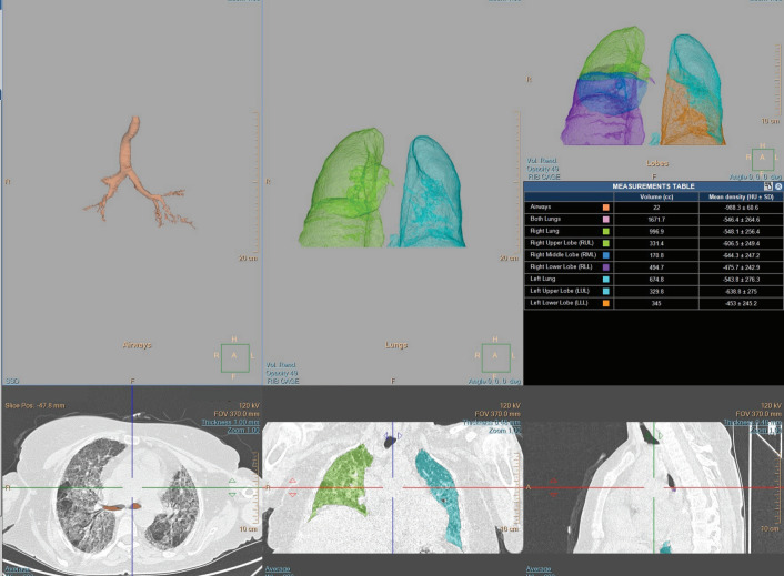
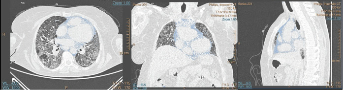
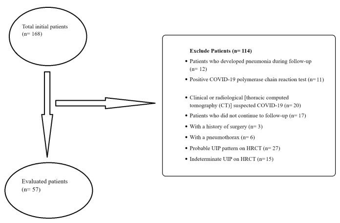
Materials and methods: The HRCT images of 57 patients diagnosed with IPF were retrospectively screened. Subcutaneous adipose tissue volume, mediastinal adipose tissue volume, and mean lung density and volume were measured at the time of diagnosis and at the 12th month. The ability of these parameters to predict progression was evaluated using the univariate and multivariate Cox regression analyses.
Results: Low mediastinal adipose tissue volume at diagnosis had a 0.991-fold effect [odds ratio (OR)= 0.991, 95% confidence interval (CI)= 0.984-0.997, p< 0.001] on progression. Low mediastinal adipose tissue volume at diagnosis had a 0.993-fold effect [odds ratio (OR)= 0.993, 95% confidence interval (CI)= 0.975-1.011, p< 0.001] and progression development at the 12th month had a 6.5-fold effect [odds ratio (OR)= 6.516, 95% confidence interval (CI)= 1.594-26.639, p< 0.009] on mortality.
Conclusion: This study indicate that the prognosis was better in those with a large mediastinal adipose tissue volume among the patients with IPF.

 求助内容:
求助内容: 应助结果提醒方式:


