{"title":"用于评估肺气肿严重程度的基于放射组学的逻辑回归模型。","authors":"Mutlu Gülbay","doi":"10.5578/tt.20239710","DOIUrl":null,"url":null,"abstract":"<p><strong>Introduction: </strong>The aim of this study is to develop a model that differentiates between the radiological patterns of severe and mild emphysema using radiomics parameters, as well as to examine the parameters included in the model.</p><p><strong>Materials and methods: </strong>Over the last 12 months, a total of 354 patients were screened based on the presence of terms such as “Fleischner”, “CLE”, and “centriacinar” in their thoracic CT reports, culminating in a study population of 82 patients. The study population was divided into Group 1 (Fleischner mild and moderate; n= 45) and Group 2 (Fleischner confluent and advanced destructive; n= 37). Volumetric segmentation was performed, focusing on the upper lobe segments of both lungs. From these segmented volumes, radiomics parameters including shape, size, first-order, and second-order features were calculated. The best model parameters were selected based on the Bayesian Information Criterion and further optimized through grid search. The final model was tested using 1000 iterations of bootstrap resampling.</p><p><strong>Results: </strong>In the training set, performance metrics were calculated with a sensitivity of 0.862, specificity of 0.870, accuracy of 0.863, and AUC of 0.910. Correspondingly, in the test set, these values were sensitivity= 0.848; specificity= 0.865; accuracy= 0.857; and AUC= 0.907.</p><p><strong>Conclusion: </strong>The logistic regression model, composed of radiomics parameters and trained on a limited number of cases, effectively differentiated between mild and severe radiological patterns of emphysema using computed tomography images.</p>","PeriodicalId":45521,"journal":{"name":"Tuberkuloz ve Toraks-Tuberculosis and Thorax","volume":"71 3","pages":"290-298"},"PeriodicalIF":0.7000,"publicationDate":"2023-09-01","publicationTypes":"Journal Article","fieldsOfStudy":null,"isOpenAccess":false,"openAccessPdf":"https://www.ncbi.nlm.nih.gov/pmc/articles/PMC10795240/pdf/","citationCount":"0","resultStr":"{\"title\":\"A radiomics-based logistic regression model for the assessment of emphysema severity.\",\"authors\":\"Mutlu Gülbay\",\"doi\":\"10.5578/tt.20239710\",\"DOIUrl\":null,\"url\":null,\"abstract\":\"<p><strong>Introduction: </strong>The aim of this study is to develop a model that differentiates between the radiological patterns of severe and mild emphysema using radiomics parameters, as well as to examine the parameters included in the model.</p><p><strong>Materials and methods: </strong>Over the last 12 months, a total of 354 patients were screened based on the presence of terms such as “Fleischner”, “CLE”, and “centriacinar” in their thoracic CT reports, culminating in a study population of 82 patients. The study population was divided into Group 1 (Fleischner mild and moderate; n= 45) and Group 2 (Fleischner confluent and advanced destructive; n= 37). Volumetric segmentation was performed, focusing on the upper lobe segments of both lungs. From these segmented volumes, radiomics parameters including shape, size, first-order, and second-order features were calculated. The best model parameters were selected based on the Bayesian Information Criterion and further optimized through grid search. The final model was tested using 1000 iterations of bootstrap resampling.</p><p><strong>Results: </strong>In the training set, performance metrics were calculated with a sensitivity of 0.862, specificity of 0.870, accuracy of 0.863, and AUC of 0.910. Correspondingly, in the test set, these values were sensitivity= 0.848; specificity= 0.865; accuracy= 0.857; and AUC= 0.907.</p><p><strong>Conclusion: </strong>The logistic regression model, composed of radiomics parameters and trained on a limited number of cases, effectively differentiated between mild and severe radiological patterns of emphysema using computed tomography images.</p>\",\"PeriodicalId\":45521,\"journal\":{\"name\":\"Tuberkuloz ve Toraks-Tuberculosis and Thorax\",\"volume\":\"71 3\",\"pages\":\"290-298\"},\"PeriodicalIF\":0.7000,\"publicationDate\":\"2023-09-01\",\"publicationTypes\":\"Journal Article\",\"fieldsOfStudy\":null,\"isOpenAccess\":false,\"openAccessPdf\":\"https://www.ncbi.nlm.nih.gov/pmc/articles/PMC10795240/pdf/\",\"citationCount\":\"0\",\"resultStr\":null,\"platform\":\"Semanticscholar\",\"paperid\":null,\"PeriodicalName\":\"Tuberkuloz ve Toraks-Tuberculosis and Thorax\",\"FirstCategoryId\":\"1085\",\"ListUrlMain\":\"https://doi.org/10.5578/tt.20239710\",\"RegionNum\":0,\"RegionCategory\":null,\"ArticlePicture\":[],\"TitleCN\":null,\"AbstractTextCN\":null,\"PMCID\":null,\"EPubDate\":\"\",\"PubModel\":\"\",\"JCR\":\"Q4\",\"JCRName\":\"RESPIRATORY SYSTEM\",\"Score\":null,\"Total\":0}","platform":"Semanticscholar","paperid":null,"PeriodicalName":"Tuberkuloz ve Toraks-Tuberculosis and Thorax","FirstCategoryId":"1085","ListUrlMain":"https://doi.org/10.5578/tt.20239710","RegionNum":0,"RegionCategory":null,"ArticlePicture":[],"TitleCN":null,"AbstractTextCN":null,"PMCID":null,"EPubDate":"","PubModel":"","JCR":"Q4","JCRName":"RESPIRATORY SYSTEM","Score":null,"Total":0}
A radiomics-based logistic regression model for the assessment of emphysema severity.
Introduction: The aim of this study is to develop a model that differentiates between the radiological patterns of severe and mild emphysema using radiomics parameters, as well as to examine the parameters included in the model.
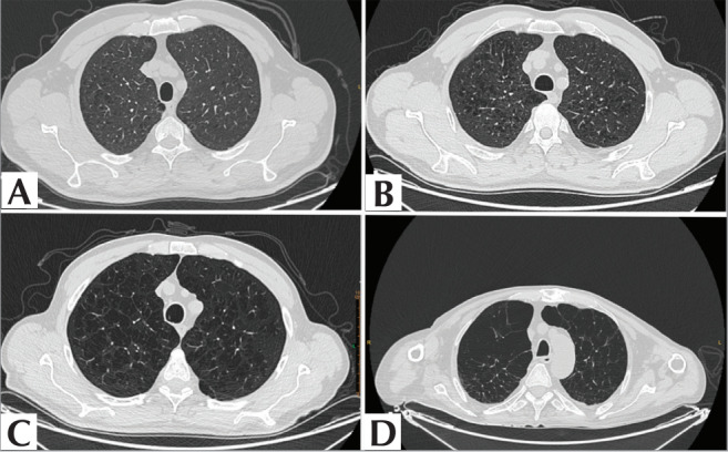
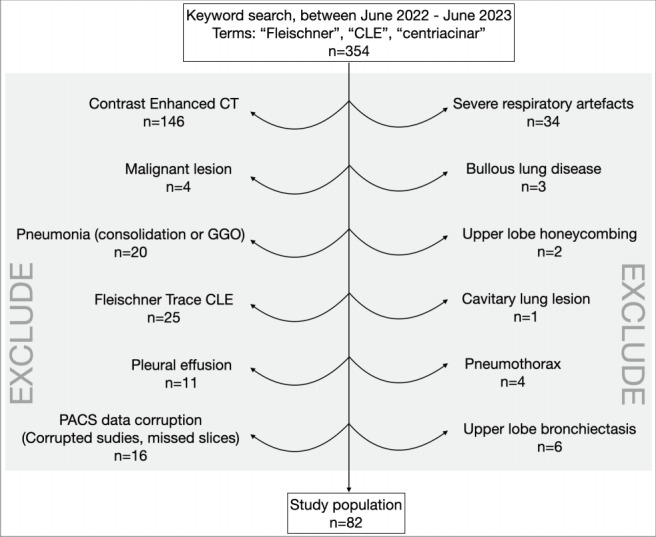
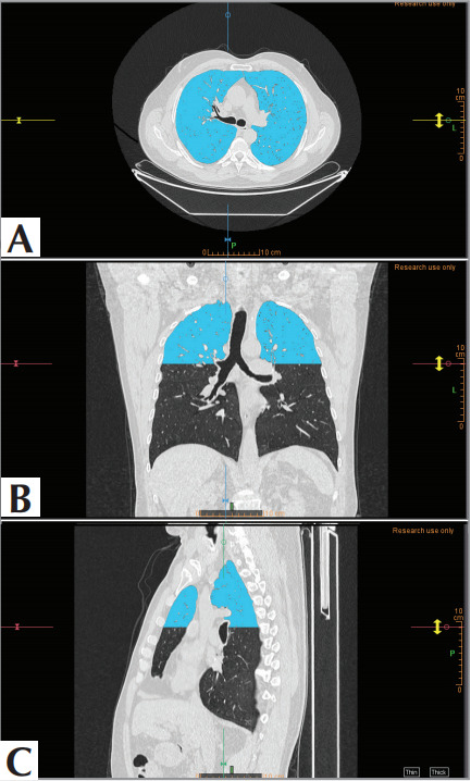
Materials and methods: Over the last 12 months, a total of 354 patients were screened based on the presence of terms such as “Fleischner”, “CLE”, and “centriacinar” in their thoracic CT reports, culminating in a study population of 82 patients. The study population was divided into Group 1 (Fleischner mild and moderate; n= 45) and Group 2 (Fleischner confluent and advanced destructive; n= 37). Volumetric segmentation was performed, focusing on the upper lobe segments of both lungs. From these segmented volumes, radiomics parameters including shape, size, first-order, and second-order features were calculated. The best model parameters were selected based on the Bayesian Information Criterion and further optimized through grid search. The final model was tested using 1000 iterations of bootstrap resampling.
Results: In the training set, performance metrics were calculated with a sensitivity of 0.862, specificity of 0.870, accuracy of 0.863, and AUC of 0.910. Correspondingly, in the test set, these values were sensitivity= 0.848; specificity= 0.865; accuracy= 0.857; and AUC= 0.907.
Conclusion: The logistic regression model, composed of radiomics parameters and trained on a limited number of cases, effectively differentiated between mild and severe radiological patterns of emphysema using computed tomography images.

 求助内容:
求助内容: 应助结果提醒方式:


