Louise Ebersberger, Fabian J Kratzer, Vanessa L Franke, Armin M Nagel, Sebastian C Niesporek, Andreas Korzowski, Mark E Ladd, Heinz-Peter Schlemmer, Daniel Paech, Tanja Platt
{"title":"在人脑神经元刺激过程中,首次在7特斯拉下实现动态氧-17(17O)磁共振成像。","authors":"Louise Ebersberger, Fabian J Kratzer, Vanessa L Franke, Armin M Nagel, Sebastian C Niesporek, Andreas Korzowski, Mark E Ladd, Heinz-Peter Schlemmer, Daniel Paech, Tanja Platt","doi":"10.1007/s10334-023-01119-6","DOIUrl":null,"url":null,"abstract":"<p><strong>Objective: </strong>First implementation of dynamic oxygen-17 (<sup>17</sup>O) MRI at 7 Tesla (T) during neuronal stimulation in the human brain.</p><p><strong>Methods: </strong>Five healthy volunteers underwent a three-phase <sup>17</sup>O gas (<sup>17</sup>O<sub>2</sub>) inhalation experiment. Combined right-side visual stimulus and right-hand finger tapping were used to achieve neuronal stimulation in the left cerebral hemisphere. Data analysis included the evaluation of the relative partial volume (PV)-corrected time evolution of absolute <sup>17</sup>O water (H<sub>2</sub><sup>17</sup>O) concentration and of the relative signal evolution without PV correction. Statistical analysis was performed using a one-tailed paired t test. Blood oxygen level-dependent (BOLD) experiments were performed to validate the stimulation paradigm.</p><p><strong>Results: </strong>The BOLD maps showed significant activity in the stimulated left visual and sensorimotor cortex compared to the non-stimulated right side. PV correction of <sup>17</sup>O MR data resulted in high signal fluctuations with a noise level of 10% due to small regions of interest (ROI), impeding further quantitative analysis. Statistical evaluation of the relative H<sub>2</sub><sup>17</sup>O signal with PV correction (p = 0.168) and without (p = 0.382) did not show significant difference between the stimulated left and non-stimulated right sensorimotor ROI.</p><p><strong>Discussion: </strong>The change of cerebral oxygen metabolism induced by sensorimotor and visual stimulation is not large enough to be reliably detected with the current setup and methodology of dynamic <sup>17</sup>O MRI at 7 T.</p>","PeriodicalId":18067,"journal":{"name":"Magnetic Resonance Materials in Physics, Biology and Medicine","volume":" ","pages":"27-38"},"PeriodicalIF":2.0000,"publicationDate":"2024-02-01","publicationTypes":"Journal Article","fieldsOfStudy":null,"isOpenAccess":false,"openAccessPdf":"https://www.ncbi.nlm.nih.gov/pmc/articles/PMC10876824/pdf/","citationCount":"0","resultStr":"{\"title\":\"First implementation of dynamic oxygen-17 (<sup>17</sup>O) magnetic resonance imaging at 7 Tesla during neuronal stimulation in the human brain.\",\"authors\":\"Louise Ebersberger, Fabian J Kratzer, Vanessa L Franke, Armin M Nagel, Sebastian C Niesporek, Andreas Korzowski, Mark E Ladd, Heinz-Peter Schlemmer, Daniel Paech, Tanja Platt\",\"doi\":\"10.1007/s10334-023-01119-6\",\"DOIUrl\":null,\"url\":null,\"abstract\":\"<p><strong>Objective: </strong>First implementation of dynamic oxygen-17 (<sup>17</sup>O) MRI at 7 Tesla (T) during neuronal stimulation in the human brain.</p><p><strong>Methods: </strong>Five healthy volunteers underwent a three-phase <sup>17</sup>O gas (<sup>17</sup>O<sub>2</sub>) inhalation experiment. Combined right-side visual stimulus and right-hand finger tapping were used to achieve neuronal stimulation in the left cerebral hemisphere. Data analysis included the evaluation of the relative partial volume (PV)-corrected time evolution of absolute <sup>17</sup>O water (H<sub>2</sub><sup>17</sup>O) concentration and of the relative signal evolution without PV correction. Statistical analysis was performed using a one-tailed paired t test. Blood oxygen level-dependent (BOLD) experiments were performed to validate the stimulation paradigm.</p><p><strong>Results: </strong>The BOLD maps showed significant activity in the stimulated left visual and sensorimotor cortex compared to the non-stimulated right side. PV correction of <sup>17</sup>O MR data resulted in high signal fluctuations with a noise level of 10% due to small regions of interest (ROI), impeding further quantitative analysis. Statistical evaluation of the relative H<sub>2</sub><sup>17</sup>O signal with PV correction (p = 0.168) and without (p = 0.382) did not show significant difference between the stimulated left and non-stimulated right sensorimotor ROI.</p><p><strong>Discussion: </strong>The change of cerebral oxygen metabolism induced by sensorimotor and visual stimulation is not large enough to be reliably detected with the current setup and methodology of dynamic <sup>17</sup>O MRI at 7 T.</p>\",\"PeriodicalId\":18067,\"journal\":{\"name\":\"Magnetic Resonance Materials in Physics, Biology and Medicine\",\"volume\":\" \",\"pages\":\"27-38\"},\"PeriodicalIF\":2.0000,\"publicationDate\":\"2024-02-01\",\"publicationTypes\":\"Journal Article\",\"fieldsOfStudy\":null,\"isOpenAccess\":false,\"openAccessPdf\":\"https://www.ncbi.nlm.nih.gov/pmc/articles/PMC10876824/pdf/\",\"citationCount\":\"0\",\"resultStr\":null,\"platform\":\"Semanticscholar\",\"paperid\":null,\"PeriodicalName\":\"Magnetic Resonance Materials in Physics, Biology and Medicine\",\"FirstCategoryId\":\"3\",\"ListUrlMain\":\"https://doi.org/10.1007/s10334-023-01119-6\",\"RegionNum\":4,\"RegionCategory\":\"医学\",\"ArticlePicture\":[],\"TitleCN\":null,\"AbstractTextCN\":null,\"PMCID\":null,\"EPubDate\":\"2023/9/22 0:00:00\",\"PubModel\":\"Epub\",\"JCR\":\"Q3\",\"JCRName\":\"RADIOLOGY, NUCLEAR MEDICINE & MEDICAL IMAGING\",\"Score\":null,\"Total\":0}","platform":"Semanticscholar","paperid":null,"PeriodicalName":"Magnetic Resonance Materials in Physics, Biology and Medicine","FirstCategoryId":"3","ListUrlMain":"https://doi.org/10.1007/s10334-023-01119-6","RegionNum":4,"RegionCategory":"医学","ArticlePicture":[],"TitleCN":null,"AbstractTextCN":null,"PMCID":null,"EPubDate":"2023/9/22 0:00:00","PubModel":"Epub","JCR":"Q3","JCRName":"RADIOLOGY, NUCLEAR MEDICINE & MEDICAL IMAGING","Score":null,"Total":0}
First implementation of dynamic oxygen-17 (17O) magnetic resonance imaging at 7 Tesla during neuronal stimulation in the human brain.
Objective: First implementation of dynamic oxygen-17 (17O) MRI at 7 Tesla (T) during neuronal stimulation in the human brain.
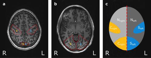
Methods: Five healthy volunteers underwent a three-phase 17O gas (17O2) inhalation experiment. Combined right-side visual stimulus and right-hand finger tapping were used to achieve neuronal stimulation in the left cerebral hemisphere. Data analysis included the evaluation of the relative partial volume (PV)-corrected time evolution of absolute 17O water (H217O) concentration and of the relative signal evolution without PV correction. Statistical analysis was performed using a one-tailed paired t test. Blood oxygen level-dependent (BOLD) experiments were performed to validate the stimulation paradigm.
Results: The BOLD maps showed significant activity in the stimulated left visual and sensorimotor cortex compared to the non-stimulated right side. PV correction of 17O MR data resulted in high signal fluctuations with a noise level of 10% due to small regions of interest (ROI), impeding further quantitative analysis. Statistical evaluation of the relative H217O signal with PV correction (p = 0.168) and without (p = 0.382) did not show significant difference between the stimulated left and non-stimulated right sensorimotor ROI.
Discussion: The change of cerebral oxygen metabolism induced by sensorimotor and visual stimulation is not large enough to be reliably detected with the current setup and methodology of dynamic 17O MRI at 7 T.
期刊介绍:
MAGMA is a multidisciplinary international journal devoted to the publication of articles on all aspects of magnetic resonance techniques and their applications in medicine and biology. MAGMA currently publishes research papers, reviews, letters to the editor, and commentaries, six times a year. The subject areas covered by MAGMA include:
advances in materials, hardware and software in magnetic resonance technology,
new developments and results in research and practical applications of magnetic resonance imaging and spectroscopy related to biology and medicine,
study of animal models and intact cells using magnetic resonance,
reports of clinical trials on humans and clinical validation of magnetic resonance protocols.

 求助内容:
求助内容: 应助结果提醒方式:


