James W. Johnson, Ben Gadomski, Kevin Labus, Holly Stewart, Brad Nelson, Howie Seim III, Dan Regan, Devin von Stade, Cambre Kelly, Phillip Horne, Ken Gall, Jeremiah Easley
{"title":"新型3D打印晶格结构钛笼在绵羊椎间融合模型中的评估。","authors":"James W. Johnson, Ben Gadomski, Kevin Labus, Holly Stewart, Brad Nelson, Howie Seim III, Dan Regan, Devin von Stade, Cambre Kelly, Phillip Horne, Ken Gall, Jeremiah Easley","doi":"10.1002/jsp2.1268","DOIUrl":null,"url":null,"abstract":"<div>\n \n \n <section>\n \n <h3> Background</h3>\n \n <p>The use of intervertebral cages within the interbody fusion setting is ubiquitous. Synthetic cages are predominantly manufactured using materials such as Ti and PEEK. With the advent of additive manufacturing techniques, it is now possible to spatially vary complex 3D geometric features within interbody devices, enabling the devices to match the stiffness of native tissue and better promote bony integration. To date, the impact of surface porosity of additively manufactured Ti interbody cages on fusion outcomes has not been investigated. Thus, the objective of this work was to determine the effect of implant endplate surface and implant body architecture of additive manufactured lattice structure titanium interbody cages on bony fusion.</p>\n </section>\n \n <section>\n \n <h3> Methods</h3>\n \n <p>Biomechanical, microcomputed tomography, static and dynamic histomorphometry, and histopathology analyses were performed on twelve functional spine units obtained from six sheep randomly allocated to body lattice or surface lattice groups.</p>\n </section>\n \n <section>\n \n <h3> Results</h3>\n \n <p>Nondestructive kinematic testing, microcomputed tomography analysis, and histomorphometry analyses of the functional spine units revealed positive fusion outcomes in both groups. These data revealed similar results in both groups, with the exception of bone-in-contact analysis, which revealed significantly improved bone-in-contact values in the body lattice group compared to the surface lattice group.</p>\n </section>\n \n <section>\n \n <h3> Conclusion</h3>\n \n <p>Both additively manufactured porous titanium cage designs resulted in increased fusion outcomes as compared to PEEK interbody cage designs as illustrated by the nondestructive kinematic motion testing, static and dynamic histomorphometry, microcomputed tomography, and histopathology analyses. While both cages provided for similar functional outcomes, these data suggest boney contact with an interbody cage may be impacted by the nature of implant porosity adjacent to the vertebral endplates.</p>\n </section>\n </div>","PeriodicalId":14876,"journal":{"name":"JOR Spine","volume":null,"pages":null},"PeriodicalIF":3.4000,"publicationDate":"2023-06-10","publicationTypes":"Journal Article","fieldsOfStudy":null,"isOpenAccess":false,"openAccessPdf":"https://ftp.ncbi.nlm.nih.gov/pub/pmc/oa_pdf/09/be/JSP2-6-e1268.PMC10540818.pdf","citationCount":"0","resultStr":"{\"title\":\"Novel 3D printed lattice structure titanium cages evaluated in an ovine model of interbody fusion\",\"authors\":\"James W. Johnson, Ben Gadomski, Kevin Labus, Holly Stewart, Brad Nelson, Howie Seim III, Dan Regan, Devin von Stade, Cambre Kelly, Phillip Horne, Ken Gall, Jeremiah Easley\",\"doi\":\"10.1002/jsp2.1268\",\"DOIUrl\":null,\"url\":null,\"abstract\":\"<div>\\n \\n \\n <section>\\n \\n <h3> Background</h3>\\n \\n <p>The use of intervertebral cages within the interbody fusion setting is ubiquitous. Synthetic cages are predominantly manufactured using materials such as Ti and PEEK. With the advent of additive manufacturing techniques, it is now possible to spatially vary complex 3D geometric features within interbody devices, enabling the devices to match the stiffness of native tissue and better promote bony integration. To date, the impact of surface porosity of additively manufactured Ti interbody cages on fusion outcomes has not been investigated. Thus, the objective of this work was to determine the effect of implant endplate surface and implant body architecture of additive manufactured lattice structure titanium interbody cages on bony fusion.</p>\\n </section>\\n \\n <section>\\n \\n <h3> Methods</h3>\\n \\n <p>Biomechanical, microcomputed tomography, static and dynamic histomorphometry, and histopathology analyses were performed on twelve functional spine units obtained from six sheep randomly allocated to body lattice or surface lattice groups.</p>\\n </section>\\n \\n <section>\\n \\n <h3> Results</h3>\\n \\n <p>Nondestructive kinematic testing, microcomputed tomography analysis, and histomorphometry analyses of the functional spine units revealed positive fusion outcomes in both groups. These data revealed similar results in both groups, with the exception of bone-in-contact analysis, which revealed significantly improved bone-in-contact values in the body lattice group compared to the surface lattice group.</p>\\n </section>\\n \\n <section>\\n \\n <h3> Conclusion</h3>\\n \\n <p>Both additively manufactured porous titanium cage designs resulted in increased fusion outcomes as compared to PEEK interbody cage designs as illustrated by the nondestructive kinematic motion testing, static and dynamic histomorphometry, microcomputed tomography, and histopathology analyses. While both cages provided for similar functional outcomes, these data suggest boney contact with an interbody cage may be impacted by the nature of implant porosity adjacent to the vertebral endplates.</p>\\n </section>\\n </div>\",\"PeriodicalId\":14876,\"journal\":{\"name\":\"JOR Spine\",\"volume\":null,\"pages\":null},\"PeriodicalIF\":3.4000,\"publicationDate\":\"2023-06-10\",\"publicationTypes\":\"Journal Article\",\"fieldsOfStudy\":null,\"isOpenAccess\":false,\"openAccessPdf\":\"https://ftp.ncbi.nlm.nih.gov/pub/pmc/oa_pdf/09/be/JSP2-6-e1268.PMC10540818.pdf\",\"citationCount\":\"0\",\"resultStr\":null,\"platform\":\"Semanticscholar\",\"paperid\":null,\"PeriodicalName\":\"JOR Spine\",\"FirstCategoryId\":\"3\",\"ListUrlMain\":\"https://onlinelibrary.wiley.com/doi/10.1002/jsp2.1268\",\"RegionNum\":3,\"RegionCategory\":\"医学\",\"ArticlePicture\":[],\"TitleCN\":null,\"AbstractTextCN\":null,\"PMCID\":null,\"EPubDate\":\"\",\"PubModel\":\"\",\"JCR\":\"Q1\",\"JCRName\":\"ORTHOPEDICS\",\"Score\":null,\"Total\":0}","platform":"Semanticscholar","paperid":null,"PeriodicalName":"JOR Spine","FirstCategoryId":"3","ListUrlMain":"https://onlinelibrary.wiley.com/doi/10.1002/jsp2.1268","RegionNum":3,"RegionCategory":"医学","ArticlePicture":[],"TitleCN":null,"AbstractTextCN":null,"PMCID":null,"EPubDate":"","PubModel":"","JCR":"Q1","JCRName":"ORTHOPEDICS","Score":null,"Total":0}
Novel 3D printed lattice structure titanium cages evaluated in an ovine model of interbody fusion
Background
The use of intervertebral cages within the interbody fusion setting is ubiquitous. Synthetic cages are predominantly manufactured using materials such as Ti and PEEK. With the advent of additive manufacturing techniques, it is now possible to spatially vary complex 3D geometric features within interbody devices, enabling the devices to match the stiffness of native tissue and better promote bony integration. To date, the impact of surface porosity of additively manufactured Ti interbody cages on fusion outcomes has not been investigated. Thus, the objective of this work was to determine the effect of implant endplate surface and implant body architecture of additive manufactured lattice structure titanium interbody cages on bony fusion.
Methods
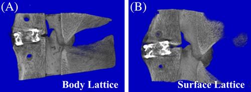
Biomechanical, microcomputed tomography, static and dynamic histomorphometry, and histopathology analyses were performed on twelve functional spine units obtained from six sheep randomly allocated to body lattice or surface lattice groups.
Results
Nondestructive kinematic testing, microcomputed tomography analysis, and histomorphometry analyses of the functional spine units revealed positive fusion outcomes in both groups. These data revealed similar results in both groups, with the exception of bone-in-contact analysis, which revealed significantly improved bone-in-contact values in the body lattice group compared to the surface lattice group.
Conclusion
Both additively manufactured porous titanium cage designs resulted in increased fusion outcomes as compared to PEEK interbody cage designs as illustrated by the nondestructive kinematic motion testing, static and dynamic histomorphometry, microcomputed tomography, and histopathology analyses. While both cages provided for similar functional outcomes, these data suggest boney contact with an interbody cage may be impacted by the nature of implant porosity adjacent to the vertebral endplates.

 求助内容:
求助内容: 应助结果提醒方式:


