下载PDF
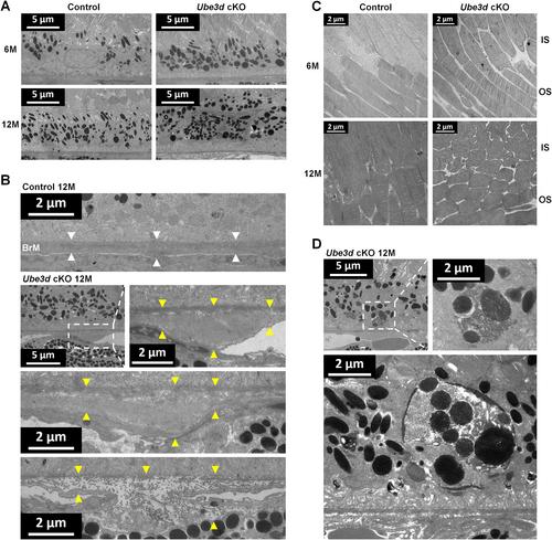
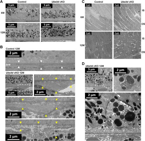
{"title":"视网膜色素上皮中Ube3d的条件性丧失加速了小鼠视网膜中与年龄相关的改变","authors":"Tianchang Tao, Ningda Xu, Jiarui Li, Mingwei Zhao, Xiaoxin Li, Lvzhen Huang","doi":"10.1002/path.6201","DOIUrl":null,"url":null,"abstract":"<p>Several studies have suggested a correlation between the ubiquitin-proteasome system (UPS) and age-related macular degeneration (AMD), with its phenotypic severity ranging from mild visual impairment to blindness, but the mechanism for UPS dysfunction contributing to disease progression is unclear. In this study, we investigated the role of ubiquitin protein ligase E3D (UBE3D) in aging and degeneration in mouse retina. Conditional knockout of <i>Ube3d</i> in the retinal pigment epithelium (RPE) of mice led to progressive and irregular fundus lesions, attenuation of the retinal vascular system, and age-associated deterioration of rod and cone responses. Simultaneously, RPE-specific <i>Ube3d</i> knockout mice also presented morphological changes similar to the histopathological characteristics of human AMD, in which a defective UPS led to RPE abnormalities such as phagocytosis or degradation of metabolites, the interaction with photoreceptor outer segment, and the transport of nutrients or waste products with choroidal capillaries via Bruch's membrane. Moreover, conditional loss of <i>Ube3d</i> resulted in aberrant molecular characterizations associated with the autophagy–lysosomal pathway, oxidative stress damage, and cell-cycle regulation, which are implicated in AMD pathology. Thus, our findings strengthen and expand the impact of UPS dysfunction on retinal pathophysiology during aging, indicating that genetic <i>Ube3d</i> deficiency in the RPE could lead to the abnormal formation of pigment deposits and secondary fundus alterations. © 2023 The Authors. <i>The Journal of Pathology</i> published by John Wiley & Sons Ltd on behalf of The Pathological Society of Great Britain and Ireland.</p>","PeriodicalId":232,"journal":{"name":"The Journal of Pathology","volume":"261 4","pages":"442-454"},"PeriodicalIF":5.6000,"publicationDate":"2023-09-29","publicationTypes":"Journal Article","fieldsOfStudy":null,"isOpenAccess":false,"openAccessPdf":"https://pathsocjournals.onlinelibrary.wiley.com/doi/epdf/10.1002/path.6201","citationCount":"0","resultStr":"{\"title\":\"Conditional loss of Ube3d in the retinal pigment epithelium accelerates age-associated alterations in the retina of mice\",\"authors\":\"Tianchang Tao, Ningda Xu, Jiarui Li, Mingwei Zhao, Xiaoxin Li, Lvzhen Huang\",\"doi\":\"10.1002/path.6201\",\"DOIUrl\":null,\"url\":null,\"abstract\":\"<p>Several studies have suggested a correlation between the ubiquitin-proteasome system (UPS) and age-related macular degeneration (AMD), with its phenotypic severity ranging from mild visual impairment to blindness, but the mechanism for UPS dysfunction contributing to disease progression is unclear. In this study, we investigated the role of ubiquitin protein ligase E3D (UBE3D) in aging and degeneration in mouse retina. Conditional knockout of <i>Ube3d</i> in the retinal pigment epithelium (RPE) of mice led to progressive and irregular fundus lesions, attenuation of the retinal vascular system, and age-associated deterioration of rod and cone responses. Simultaneously, RPE-specific <i>Ube3d</i> knockout mice also presented morphological changes similar to the histopathological characteristics of human AMD, in which a defective UPS led to RPE abnormalities such as phagocytosis or degradation of metabolites, the interaction with photoreceptor outer segment, and the transport of nutrients or waste products with choroidal capillaries via Bruch's membrane. Moreover, conditional loss of <i>Ube3d</i> resulted in aberrant molecular characterizations associated with the autophagy–lysosomal pathway, oxidative stress damage, and cell-cycle regulation, which are implicated in AMD pathology. Thus, our findings strengthen and expand the impact of UPS dysfunction on retinal pathophysiology during aging, indicating that genetic <i>Ube3d</i> deficiency in the RPE could lead to the abnormal formation of pigment deposits and secondary fundus alterations. © 2023 The Authors. <i>The Journal of Pathology</i> published by John Wiley & Sons Ltd on behalf of The Pathological Society of Great Britain and Ireland.</p>\",\"PeriodicalId\":232,\"journal\":{\"name\":\"The Journal of Pathology\",\"volume\":\"261 4\",\"pages\":\"442-454\"},\"PeriodicalIF\":5.6000,\"publicationDate\":\"2023-09-29\",\"publicationTypes\":\"Journal Article\",\"fieldsOfStudy\":null,\"isOpenAccess\":false,\"openAccessPdf\":\"https://pathsocjournals.onlinelibrary.wiley.com/doi/epdf/10.1002/path.6201\",\"citationCount\":\"0\",\"resultStr\":null,\"platform\":\"Semanticscholar\",\"paperid\":null,\"PeriodicalName\":\"The Journal of Pathology\",\"FirstCategoryId\":\"3\",\"ListUrlMain\":\"https://onlinelibrary.wiley.com/doi/10.1002/path.6201\",\"RegionNum\":2,\"RegionCategory\":\"医学\",\"ArticlePicture\":[],\"TitleCN\":null,\"AbstractTextCN\":null,\"PMCID\":null,\"EPubDate\":\"\",\"PubModel\":\"\",\"JCR\":\"Q1\",\"JCRName\":\"ONCOLOGY\",\"Score\":null,\"Total\":0}","platform":"Semanticscholar","paperid":null,"PeriodicalName":"The Journal of Pathology","FirstCategoryId":"3","ListUrlMain":"https://onlinelibrary.wiley.com/doi/10.1002/path.6201","RegionNum":2,"RegionCategory":"医学","ArticlePicture":[],"TitleCN":null,"AbstractTextCN":null,"PMCID":null,"EPubDate":"","PubModel":"","JCR":"Q1","JCRName":"ONCOLOGY","Score":null,"Total":0}
引用次数: 0
引用
批量引用


 求助内容:
求助内容: 应助结果提醒方式:


