上颌中央切牙的牙本质壁厚度测量与牙根发育阶段的关系:一项初步研究。

IF 1.4 Q3 DENTISTRY, ORAL SURGERY & MEDICINE
Petra Bučević Sojčić, Jasna Leder Horina, Tanja Jurčević Lulić, Nina Bočkaj, Hrvoje Jurić
{"title":"上颌中央切牙的牙本质壁厚度测量与牙根发育阶段的关系:一项初步研究。","authors":"Petra Bučević Sojčić, Jasna Leder Horina, Tanja Jurčević Lulić, Nina Bočkaj, Hrvoje Jurić","doi":"10.15644/asc57/3/1","DOIUrl":null,"url":null,"abstract":"Objective The aim of this study was to determine the average dentin wall thickness (DWT) of the maxillary central incisor (MCI) required for performing finite element analysis (FEA) models of root development. Material and methods A total of 137 intraoral periapical radiographs of MCI in children aged 7 to 11 years were examined and then classified into 5 groups according to root development stages, which included 1/2 of root development (S1), 3/4 of root development (S2), more than 3/4 of root development (S3), complete development with wide-open apex (S4) and complete development with closed apex (S5). DWT was measured at three reference (horizontal) lines: at a distance of 1 mm from the apex (M), 4 mm from the apex (L) and at the cervical line (K). The distal dentin wall thickness (M1, L1, and K1), the pulp thickness (M2, L2, and K2), the mesial dentin wall thickness (M3, L3, and K3), and the apex thickness (N) were measured using the diagnostic software Soredex Scanora 5.1.2.4. Statistical analysis compared the values of the parameters K, L, and M between developmental stages (multivariate ANOVA) and the linear correlations between the parameters (Pearson's correlation analysis). All analyses were performed at significance level α = 0.05. Results There were statistically significant differences between the developmental stages for parameters L and M, while no significant differences were found for parameter K. Most of the correlations between the parameters were statistically significant, with the values of the Pearson correlation coefficient R > 0.6 considered practically significant. All parameters on the same reference line for distal and mesial dentin wall thickness and for pulp thickness correlated well with each other (R = 0.46 – 0.68), but there was no statistically significant correlation with total root thickness on the same reference line (parameters K, L, or M), except for parameter K3 (R = 0.42). Conclusion Despite the limitations of this study, the mean values of the selected parameters for the 5 groups of developmental stages of the maxillary central incisor could be used to model dentin wall thickness using finite element analysis.","PeriodicalId":7154,"journal":{"name":"Acta Stomatologica Croatica","volume":"57 3","pages":"206-215"},"PeriodicalIF":1.4000,"publicationDate":"2023-09-01","publicationTypes":"Journal Article","fieldsOfStudy":null,"isOpenAccess":false,"openAccessPdf":"https://ftp.ncbi.nlm.nih.gov/pub/pmc/oa_pdf/50/28/ASC_57(3)_206-215.PMC10557114.pdf","citationCount":"0","resultStr":"{\"title\":\"Measurement of the Dentin Wall Thickness of the Maxillary Central Incisor in Relation to the Stage of Root Development: A Pilot Study.\",\"authors\":\"Petra Bučević Sojčić, Jasna Leder Horina, Tanja Jurčević Lulić, Nina Bočkaj, Hrvoje Jurić\",\"doi\":\"10.15644/asc57/3/1\",\"DOIUrl\":null,\"url\":null,\"abstract\":\"Objective The aim of this study was to determine the average dentin wall thickness (DWT) of the maxillary central incisor (MCI) required for performing finite element analysis (FEA) models of root development. Material and methods A total of 137 intraoral periapical radiographs of MCI in children aged 7 to 11 years were examined and then classified into 5 groups according to root development stages, which included 1/2 of root development (S1), 3/4 of root development (S2), more than 3/4 of root development (S3), complete development with wide-open apex (S4) and complete development with closed apex (S5). DWT was measured at three reference (horizontal) lines: at a distance of 1 mm from the apex (M), 4 mm from the apex (L) and at the cervical line (K). The distal dentin wall thickness (M1, L1, and K1), the pulp thickness (M2, L2, and K2), the mesial dentin wall thickness (M3, L3, and K3), and the apex thickness (N) were measured using the diagnostic software Soredex Scanora 5.1.2.4. Statistical analysis compared the values of the parameters K, L, and M between developmental stages (multivariate ANOVA) and the linear correlations between the parameters (Pearson's correlation analysis). All analyses were performed at significance level α = 0.05. Results There were statistically significant differences between the developmental stages for parameters L and M, while no significant differences were found for parameter K. Most of the correlations between the parameters were statistically significant, with the values of the Pearson correlation coefficient R > 0.6 considered practically significant. All parameters on the same reference line for distal and mesial dentin wall thickness and for pulp thickness correlated well with each other (R = 0.46 – 0.68), but there was no statistically significant correlation with total root thickness on the same reference line (parameters K, L, or M), except for parameter K3 (R = 0.42). Conclusion Despite the limitations of this study, the mean values of the selected parameters for the 5 groups of developmental stages of the maxillary central incisor could be used to model dentin wall thickness using finite element analysis.\",\"PeriodicalId\":7154,\"journal\":{\"name\":\"Acta Stomatologica Croatica\",\"volume\":\"57 3\",\"pages\":\"206-215\"},\"PeriodicalIF\":1.4000,\"publicationDate\":\"2023-09-01\",\"publicationTypes\":\"Journal Article\",\"fieldsOfStudy\":null,\"isOpenAccess\":false,\"openAccessPdf\":\"https://ftp.ncbi.nlm.nih.gov/pub/pmc/oa_pdf/50/28/ASC_57(3)_206-215.PMC10557114.pdf\",\"citationCount\":\"0\",\"resultStr\":null,\"platform\":\"Semanticscholar\",\"paperid\":null,\"PeriodicalName\":\"Acta Stomatologica Croatica\",\"FirstCategoryId\":\"1085\",\"ListUrlMain\":\"https://doi.org/10.15644/asc57/3/1\",\"RegionNum\":0,\"RegionCategory\":null,\"ArticlePicture\":[],\"TitleCN\":null,\"AbstractTextCN\":null,\"PMCID\":null,\"EPubDate\":\"\",\"PubModel\":\"\",\"JCR\":\"Q3\",\"JCRName\":\"DENTISTRY, ORAL SURGERY & MEDICINE\",\"Score\":null,\"Total\":0}","platform":"Semanticscholar","paperid":null,"PeriodicalName":"Acta Stomatologica Croatica","FirstCategoryId":"1085","ListUrlMain":"https://doi.org/10.15644/asc57/3/1","RegionNum":0,"RegionCategory":null,"ArticlePicture":[],"TitleCN":null,"AbstractTextCN":null,"PMCID":null,"EPubDate":"","PubModel":"","JCR":"Q3","JCRName":"DENTISTRY, ORAL SURGERY & MEDICINE","Score":null,"Total":0}
引用次数: 0

摘要

目的:本研究的目的是确定上颌中切牙(MCI)的平均牙本质壁厚(DWT),以进行牙根发育的有限元分析(FEA)模型。材料与方法:对7~11岁儿童口腔内MCI的137张根尖周X线片进行检查,然后根据牙根发育阶段分为5组,包括1/2牙根发育(S1)、3/4牙根发育(S2)、3/4以上牙根发育(S3),完全发育具有宽的开放顶点(S4)和完全发育具有闭合顶点(S5)。在三条参考(水平)线上测量DWT:距离顶点(M)1mm、距离顶点(L)4mm和宫颈线(K)。使用诊断软件Soredex Scanora 5.1.2.4测量远端牙本质壁厚(M1、L1和K1)、牙髓厚度(M2、L2和K2)、近中牙本质壁厚度(M3、L3和K3)和根尖厚度(N)。统计分析比较了发育阶段之间的参数K、L和M的值(多变量方差分析)和参数之间的线性相关性(Pearson相关分析)。所有分析均在显著性水平α=0.05下进行。结果:参数L和M在发育阶段之间存在统计学显著差异,而参数K没有发现显著差异。大多数参数之间的相关性具有统计学显著性,Pearson相关系数R>0.6的值被认为实际上是显著的。在同一参考线上,远端和近端牙本质壁厚度以及牙髓厚度的所有参数相互关联良好(R=0.46-0.68),但除了参数K3(R=0.42)外,与同一参考线上的总根厚度(参数K、L或M)没有统计学显著相关性。结论:尽管本研究存在局限性,上颌中切牙5组发育阶段所选参数的平均值可用于有限元分析牙本质壁厚模型。
本文章由计算机程序翻译,如有差异,请以英文原文为准。

Measurement of the Dentin Wall Thickness of the Maxillary Central Incisor in Relation to the Stage of Root Development: A Pilot Study.

Measurement of the Dentin Wall Thickness of the Maxillary Central Incisor in Relation to the Stage of Root Development: A Pilot Study.

Measurement of the Dentin Wall Thickness of the Maxillary Central Incisor in Relation to the Stage of Root Development: A Pilot Study.

Measurement of the Dentin Wall Thickness of the Maxillary Central Incisor in Relation to the Stage of Root Development: A Pilot Study.
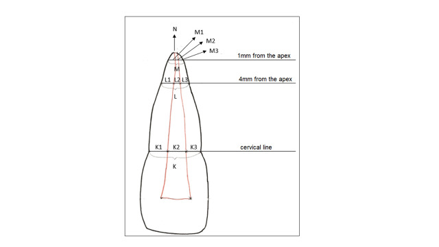
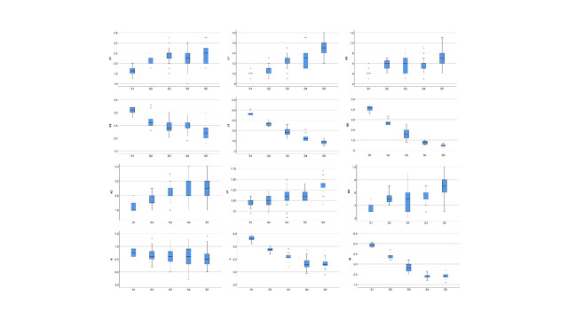
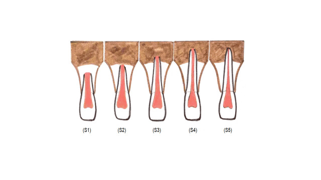
Objective The aim of this study was to determine the average dentin wall thickness (DWT) of the maxillary central incisor (MCI) required for performing finite element analysis (FEA) models of root development. Material and methods A total of 137 intraoral periapical radiographs of MCI in children aged 7 to 11 years were examined and then classified into 5 groups according to root development stages, which included 1/2 of root development (S1), 3/4 of root development (S2), more than 3/4 of root development (S3), complete development with wide-open apex (S4) and complete development with closed apex (S5). DWT was measured at three reference (horizontal) lines: at a distance of 1 mm from the apex (M), 4 mm from the apex (L) and at the cervical line (K). The distal dentin wall thickness (M1, L1, and K1), the pulp thickness (M2, L2, and K2), the mesial dentin wall thickness (M3, L3, and K3), and the apex thickness (N) were measured using the diagnostic software Soredex Scanora 5.1.2.4. Statistical analysis compared the values of the parameters K, L, and M between developmental stages (multivariate ANOVA) and the linear correlations between the parameters (Pearson's correlation analysis). All analyses were performed at significance level α = 0.05. Results There were statistically significant differences between the developmental stages for parameters L and M, while no significant differences were found for parameter K. Most of the correlations between the parameters were statistically significant, with the values of the Pearson correlation coefficient R > 0.6 considered practically significant. All parameters on the same reference line for distal and mesial dentin wall thickness and for pulp thickness correlated well with each other (R = 0.46 – 0.68), but there was no statistically significant correlation with total root thickness on the same reference line (parameters K, L, or M), except for parameter K3 (R = 0.42). Conclusion Despite the limitations of this study, the mean values of the selected parameters for the 5 groups of developmental stages of the maxillary central incisor could be used to model dentin wall thickness using finite element analysis.
求助全文
通过发布文献求助,成功后即可免费获取论文全文。 去求助
来源期刊
Acta Stomatologica Croatica
Acta Stomatologica Croatica DENTISTRY, ORAL SURGERY & MEDICINE-
CiteScore
2.40
自引率
28.60%
发文量
32
审稿时长
12 weeks
期刊介绍: The Acta Stomatologica Croatica (ASCRO) is a leading scientific non-profit journal in the field of dental, oral and cranio-facial sciences during the past 44 years in Croatia. ASCRO publishes original scientific and clinical papers, preliminary communications, case reports, book reviews, letters to the editor and news. Review articles are published by invitation from the Editor-in-Chief by acclaimed professionals in distinct fields of dental medicine. All manuscripts are subjected to peer review process.
×
引用
GB/T 7714-2015
复制
MLA
复制
APA
复制
导出至
BibTeX EndNote RefMan NoteFirst NoteExpress
×
提示
您的信息不完整,为了账户安全,请先补充。
现在去补充
×
提示
您因"违规操作"
具体请查看互助需知
我知道了
×
提示
确定
请完成安全验证×
copy
已复制链接
快去分享给好友吧!
我知道了
右上角分享
点击右上角分享
0
联系我们:info@booksci.cn Book学术提供免费学术资源搜索服务,方便国内外学者检索中英文文献。致力于提供最便捷和优质的服务体验。 Copyright © 2023 布克学术 All rights reserved.
京ICP备2023020795号-1
ghs 京公网安备 11010802042870号
Book学术文献互助
Book学术文献互助群
群 号:481959085
Book学术官方微信