Ana Paula F Trombone, Celio L Silva, Luciana P Almeida, Rogerio S Rosada, Karla M Lima, Constance Oliver, Maria C Jamur, Arlete A M Coelho-Castelo
{"title":"DNA-Hsp65/ tdm负载PLGA微球的组织分布及吞噬细胞的摄取。","authors":"Ana Paula F Trombone, Celio L Silva, Luciana P Almeida, Rogerio S Rosada, Karla M Lima, Constance Oliver, Maria C Jamur, Arlete A M Coelho-Castelo","doi":"10.1186/1479-0556-5-9","DOIUrl":null,"url":null,"abstract":"<p><p>This study aimed to demonstrate that microspheres, used as delivery vehicle of DNA-Hsp65/TDM [plasmid DNA encoding heat shock protein 65 (Hsp65) coencapsulated with trehalose dimycolate (TDM) into PLGA microspheres], are widely spread among several organs after intramuscular administration in BALB/c mice. In general, we showed that these particles were phagocytosed by antigen presenting cells, such as macrophages and dendritic cells. Besides, it was demonstrated herein that draining lymph node cells presented a significant increase in the number of cells expressing costimulatory molecules (CD80 and CD86) and MHC class II, and also that the administration of the DNA-Hsp65/TDM and vector/TDM formulations resulted in the up-regulation of CD80, CD86 and MHC class II expression when compared to control formulations (vector/TDM and empty). Regarding the intracellular trafficking we observed that following phagocytosis, the microspheres were not found in the late endosomes and/or lysosomes, until 15 days after internalization, and we suggest that these constructions were hydrolysed in early compartments. Overall, these data expand our knowledge on PLGA [poly (lactic-co-glycolic acid)] microspheres as gene carriers in vaccination strategies, as well as open perspectives for their potential use in clinical practice.</p>","PeriodicalId":12596,"journal":{"name":"Genetic Vaccines and Therapy","volume":" ","pages":"9"},"PeriodicalIF":0.0000,"publicationDate":"2007-09-20","publicationTypes":"Journal Article","fieldsOfStudy":null,"isOpenAccess":false,"openAccessPdf":"https://sci-hub-pdf.com/10.1186/1479-0556-5-9","citationCount":"15","resultStr":"{\"title\":\"Tissue distribution of DNA-Hsp65/TDM-loaded PLGA microspheres and uptake by phagocytic cells.\",\"authors\":\"Ana Paula F Trombone, Celio L Silva, Luciana P Almeida, Rogerio S Rosada, Karla M Lima, Constance Oliver, Maria C Jamur, Arlete A M Coelho-Castelo\",\"doi\":\"10.1186/1479-0556-5-9\",\"DOIUrl\":null,\"url\":null,\"abstract\":\"<p><p>This study aimed to demonstrate that microspheres, used as delivery vehicle of DNA-Hsp65/TDM [plasmid DNA encoding heat shock protein 65 (Hsp65) coencapsulated with trehalose dimycolate (TDM) into PLGA microspheres], are widely spread among several organs after intramuscular administration in BALB/c mice. In general, we showed that these particles were phagocytosed by antigen presenting cells, such as macrophages and dendritic cells. Besides, it was demonstrated herein that draining lymph node cells presented a significant increase in the number of cells expressing costimulatory molecules (CD80 and CD86) and MHC class II, and also that the administration of the DNA-Hsp65/TDM and vector/TDM formulations resulted in the up-regulation of CD80, CD86 and MHC class II expression when compared to control formulations (vector/TDM and empty). Regarding the intracellular trafficking we observed that following phagocytosis, the microspheres were not found in the late endosomes and/or lysosomes, until 15 days after internalization, and we suggest that these constructions were hydrolysed in early compartments. Overall, these data expand our knowledge on PLGA [poly (lactic-co-glycolic acid)] microspheres as gene carriers in vaccination strategies, as well as open perspectives for their potential use in clinical practice.</p>\",\"PeriodicalId\":12596,\"journal\":{\"name\":\"Genetic Vaccines and Therapy\",\"volume\":\" \",\"pages\":\"9\"},\"PeriodicalIF\":0.0000,\"publicationDate\":\"2007-09-20\",\"publicationTypes\":\"Journal Article\",\"fieldsOfStudy\":null,\"isOpenAccess\":false,\"openAccessPdf\":\"https://sci-hub-pdf.com/10.1186/1479-0556-5-9\",\"citationCount\":\"15\",\"resultStr\":null,\"platform\":\"Semanticscholar\",\"paperid\":null,\"PeriodicalName\":\"Genetic Vaccines and Therapy\",\"FirstCategoryId\":\"1085\",\"ListUrlMain\":\"https://doi.org/10.1186/1479-0556-5-9\",\"RegionNum\":0,\"RegionCategory\":null,\"ArticlePicture\":[],\"TitleCN\":null,\"AbstractTextCN\":null,\"PMCID\":null,\"EPubDate\":\"\",\"PubModel\":\"\",\"JCR\":\"\",\"JCRName\":\"\",\"Score\":null,\"Total\":0}","platform":"Semanticscholar","paperid":null,"PeriodicalName":"Genetic Vaccines and Therapy","FirstCategoryId":"1085","ListUrlMain":"https://doi.org/10.1186/1479-0556-5-9","RegionNum":0,"RegionCategory":null,"ArticlePicture":[],"TitleCN":null,"AbstractTextCN":null,"PMCID":null,"EPubDate":"","PubModel":"","JCR":"","JCRName":"","Score":null,"Total":0}
Tissue distribution of DNA-Hsp65/TDM-loaded PLGA microspheres and uptake by phagocytic cells.
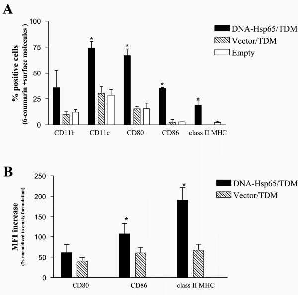
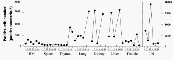
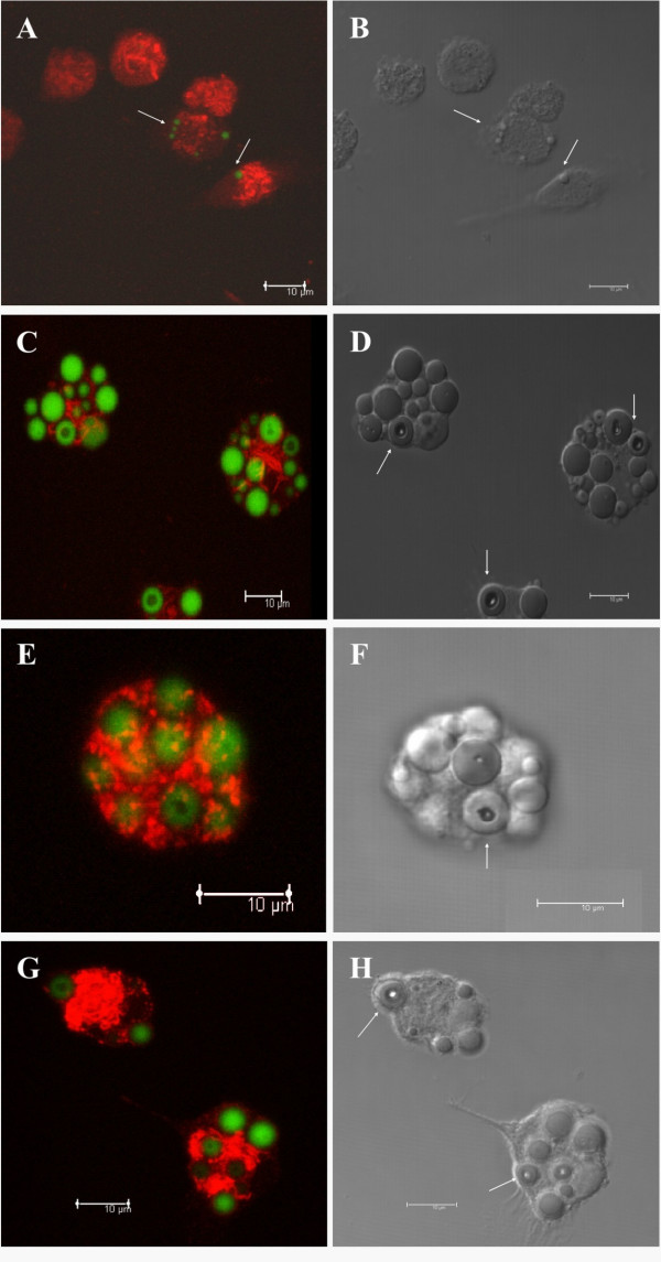
This study aimed to demonstrate that microspheres, used as delivery vehicle of DNA-Hsp65/TDM [plasmid DNA encoding heat shock protein 65 (Hsp65) coencapsulated with trehalose dimycolate (TDM) into PLGA microspheres], are widely spread among several organs after intramuscular administration in BALB/c mice. In general, we showed that these particles were phagocytosed by antigen presenting cells, such as macrophages and dendritic cells. Besides, it was demonstrated herein that draining lymph node cells presented a significant increase in the number of cells expressing costimulatory molecules (CD80 and CD86) and MHC class II, and also that the administration of the DNA-Hsp65/TDM and vector/TDM formulations resulted in the up-regulation of CD80, CD86 and MHC class II expression when compared to control formulations (vector/TDM and empty). Regarding the intracellular trafficking we observed that following phagocytosis, the microspheres were not found in the late endosomes and/or lysosomes, until 15 days after internalization, and we suggest that these constructions were hydrolysed in early compartments. Overall, these data expand our knowledge on PLGA [poly (lactic-co-glycolic acid)] microspheres as gene carriers in vaccination strategies, as well as open perspectives for their potential use in clinical practice.

 求助内容:
求助内容: 应助结果提醒方式:


