Gulsah Yildirim, Hakki Muammer Karakas, Baris Yilmaz
{"title":"骨样骨瘤的非冷却微波消融:解决老问题的新方法。","authors":"Gulsah Yildirim, Hakki Muammer Karakas, Baris Yilmaz","doi":"10.14744/nci.2021.26675","DOIUrl":null,"url":null,"abstract":"<p><strong>Objective: </strong>This study aims to evaluate the technical and clinical success of uncooled microwave ablation (MWA) in the treatment of osteoid osteoma with two-dimensional fluoroscopy guidance in the operating room.</p><p><strong>Methods: </strong>The clinical and imaging data of 9 patients were retrospectively evaluated. Mean patient age was 14.55 years. The mean size and volume of the lesions were 17.2 × 10.8 × 8.0 mm and the mean nidus size was 6.86±2.05 mm on computed tomography. MWA was performed with uncooled probe in operating room and in sterile conditions. Numerical pain score was recorded before the procedure, the day after, and at 1, 3 months after the procedure.</p><p><strong>Results: </strong>Clinical and technical success was achieved in 100% of patients. The mean volume of MWA-induced necrosis was 20.8 × 12.8 × 10.7 mm, peripheral scar thickness was 3.5±0.75 mm, and none of the patients had nidus enhancement on first month follow-up magnetic resonance imaging. Fluoroscopic guidance was conducted under digital c-arm. Patients received four to 12 spot films (mean: 6.6 kVp, 2.66 mAs) over the lower extremity. Mean radiation exposure to the skin due to imaging was 0.02 mGy per patient per procedure. The dose area product-the total amount of radiation deliverable to the patient was 0.75±0.32 Gy.cm<sup>2</sup>.</p><p><strong>Conclusion: </strong>This study demonstrated the effectiveness and the safety of the uncooled MWA in osteoid osteoma. The technique may effectively be used in operating room under c-arm fluoroscopy. Such hybrid approach may ensure sterility, anesthetic safety, and lower radiation dose to patients.</p>","PeriodicalId":19164,"journal":{"name":"Northern Clinics of Istanbul","volume":"9 5","pages":"524-529"},"PeriodicalIF":0.9000,"publicationDate":"2022-10-27","publicationTypes":"Journal Article","fieldsOfStudy":null,"isOpenAccess":false,"openAccessPdf":"https://ftp.ncbi.nlm.nih.gov/pub/pmc/oa_pdf/89/6d/NCI-9-524.PMC9677055.pdf","citationCount":"1","resultStr":"{\"title\":\"Uncooled microwave ablation of osteoid osteoma: New approaches to an old problem.\",\"authors\":\"Gulsah Yildirim, Hakki Muammer Karakas, Baris Yilmaz\",\"doi\":\"10.14744/nci.2021.26675\",\"DOIUrl\":null,\"url\":null,\"abstract\":\"<p><strong>Objective: </strong>This study aims to evaluate the technical and clinical success of uncooled microwave ablation (MWA) in the treatment of osteoid osteoma with two-dimensional fluoroscopy guidance in the operating room.</p><p><strong>Methods: </strong>The clinical and imaging data of 9 patients were retrospectively evaluated. Mean patient age was 14.55 years. The mean size and volume of the lesions were 17.2 × 10.8 × 8.0 mm and the mean nidus size was 6.86±2.05 mm on computed tomography. MWA was performed with uncooled probe in operating room and in sterile conditions. Numerical pain score was recorded before the procedure, the day after, and at 1, 3 months after the procedure.</p><p><strong>Results: </strong>Clinical and technical success was achieved in 100% of patients. The mean volume of MWA-induced necrosis was 20.8 × 12.8 × 10.7 mm, peripheral scar thickness was 3.5±0.75 mm, and none of the patients had nidus enhancement on first month follow-up magnetic resonance imaging. Fluoroscopic guidance was conducted under digital c-arm. Patients received four to 12 spot films (mean: 6.6 kVp, 2.66 mAs) over the lower extremity. Mean radiation exposure to the skin due to imaging was 0.02 mGy per patient per procedure. The dose area product-the total amount of radiation deliverable to the patient was 0.75±0.32 Gy.cm<sup>2</sup>.</p><p><strong>Conclusion: </strong>This study demonstrated the effectiveness and the safety of the uncooled MWA in osteoid osteoma. The technique may effectively be used in operating room under c-arm fluoroscopy. Such hybrid approach may ensure sterility, anesthetic safety, and lower radiation dose to patients.</p>\",\"PeriodicalId\":19164,\"journal\":{\"name\":\"Northern Clinics of Istanbul\",\"volume\":\"9 5\",\"pages\":\"524-529\"},\"PeriodicalIF\":0.9000,\"publicationDate\":\"2022-10-27\",\"publicationTypes\":\"Journal Article\",\"fieldsOfStudy\":null,\"isOpenAccess\":false,\"openAccessPdf\":\"https://ftp.ncbi.nlm.nih.gov/pub/pmc/oa_pdf/89/6d/NCI-9-524.PMC9677055.pdf\",\"citationCount\":\"1\",\"resultStr\":null,\"platform\":\"Semanticscholar\",\"paperid\":null,\"PeriodicalName\":\"Northern Clinics of Istanbul\",\"FirstCategoryId\":\"1085\",\"ListUrlMain\":\"https://doi.org/10.14744/nci.2021.26675\",\"RegionNum\":0,\"RegionCategory\":null,\"ArticlePicture\":[],\"TitleCN\":null,\"AbstractTextCN\":null,\"PMCID\":null,\"EPubDate\":\"2022/1/1 0:00:00\",\"PubModel\":\"eCollection\",\"JCR\":\"Q3\",\"JCRName\":\"MEDICINE, GENERAL & INTERNAL\",\"Score\":null,\"Total\":0}","platform":"Semanticscholar","paperid":null,"PeriodicalName":"Northern Clinics of Istanbul","FirstCategoryId":"1085","ListUrlMain":"https://doi.org/10.14744/nci.2021.26675","RegionNum":0,"RegionCategory":null,"ArticlePicture":[],"TitleCN":null,"AbstractTextCN":null,"PMCID":null,"EPubDate":"2022/1/1 0:00:00","PubModel":"eCollection","JCR":"Q3","JCRName":"MEDICINE, GENERAL & INTERNAL","Score":null,"Total":0}
Uncooled microwave ablation of osteoid osteoma: New approaches to an old problem.
Objective: This study aims to evaluate the technical and clinical success of uncooled microwave ablation (MWA) in the treatment of osteoid osteoma with two-dimensional fluoroscopy guidance in the operating room.
Methods: The clinical and imaging data of 9 patients were retrospectively evaluated. Mean patient age was 14.55 years. The mean size and volume of the lesions were 17.2 × 10.8 × 8.0 mm and the mean nidus size was 6.86±2.05 mm on computed tomography. MWA was performed with uncooled probe in operating room and in sterile conditions. Numerical pain score was recorded before the procedure, the day after, and at 1, 3 months after the procedure.
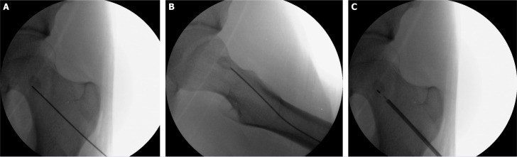
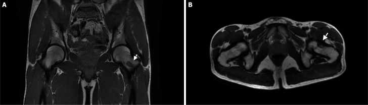
Results: Clinical and technical success was achieved in 100% of patients. The mean volume of MWA-induced necrosis was 20.8 × 12.8 × 10.7 mm, peripheral scar thickness was 3.5±0.75 mm, and none of the patients had nidus enhancement on first month follow-up magnetic resonance imaging. Fluoroscopic guidance was conducted under digital c-arm. Patients received four to 12 spot films (mean: 6.6 kVp, 2.66 mAs) over the lower extremity. Mean radiation exposure to the skin due to imaging was 0.02 mGy per patient per procedure. The dose area product-the total amount of radiation deliverable to the patient was 0.75±0.32 Gy.cm2.
Conclusion: This study demonstrated the effectiveness and the safety of the uncooled MWA in osteoid osteoma. The technique may effectively be used in operating room under c-arm fluoroscopy. Such hybrid approach may ensure sterility, anesthetic safety, and lower radiation dose to patients.

 求助内容:
求助内容: 应助结果提醒方式:


