Molecular VisionPub Date : 2022-08-07eCollection Date: 2022-01-01
Bindu Kodati, Nolan R McGrady, Hayden B Jefferies, Dorota L Stankowska, Raghu R Krishnamoorthy
{"title":"口服双重ETA/ETB受体拮抗剂促进青光眼啮齿动物模型的神经保护。","authors":"Bindu Kodati, Nolan R McGrady, Hayden B Jefferies, Dorota L Stankowska, Raghu R Krishnamoorthy","doi":"","DOIUrl":null,"url":null,"abstract":"<p><strong>Purpose: </strong>Glaucoma is a neurodegenerative disease associated with elevated intraocular pressure and characterized by optic nerve axonal degeneration, cupping of the optic disc, and loss of retinal ganglion cells (RGCs). The endothelin (ET) system of vasoactive peptides (ET-1, ET-2, ET-3) and their G-protein coupled receptors (ET<sub>A</sub> and ET<sub>B</sub> receptors) have been shown to contribute to the pathophysiology of glaucoma. The purpose of this study was to determine whether administration of the endothelin receptor antagonist macitentan was neuroprotective to RGCs and optic nerve axons when administered after the onset of intraocular pressure (IOP) elevation in ocular hypertensive rats.</p><p><strong>Methods: </strong>Male and female Brown Norway rats were subjected to the Morrison model of ocular hypertension by injection of hypertonic saline through the episcleral veins. Following IOP elevation, macitentan (5 mg/kg body wt) was administered orally 3 days per week, and rats with IOP elevation were maintained for 4 weeks. RGC function was determined by pattern electroretinography (PERG) at 2 and 4 weeks post-IOP elevation. Rats were euthanized by approved humane methods, and retinal flat mounts were generated and immunostained for the RGC-selective marker Brn3a. PPD-stained optic nerve sections were imaged by confocal microscopy. RGC and axon counts were conducted in a masked manner and compared between the treatment groups.</p><p><strong>Results: </strong>Significant protection against loss of RGCs and optic nerve axons was found following oral administration of macitentan in rats with elevated IOP. In addition, a protective trend for RGC function, as measured by pattern ERG analysis, was evident following macitentan treatment.</p><p><strong>Conclusions: </strong>Macitentan treatment had a neuroprotective effect on RGCs and their axons, independent of its IOP-lowering effect, suggesting that macitentan may complement existing treatments to prevent neurodegeneration during ocular hypertension. The findings presented have implications for the use of macitentan as an oral formulation to promote neuroprotection in glaucoma patients.</p>","PeriodicalId":18866,"journal":{"name":"Molecular Vision","volume":" ","pages":"165-177"},"PeriodicalIF":1.4000,"publicationDate":"2022-08-07","publicationTypes":"Journal Article","fieldsOfStudy":null,"isOpenAccess":false,"openAccessPdf":"https://ftp.ncbi.nlm.nih.gov/pub/pmc/oa_pdf/ee/13/mv-v28-165.PMC9491150.pdf","citationCount":"0","resultStr":"{\"title\":\"Oral administration of a dual ET<sub>A</sub>/ET<sub>B</sub> receptor antagonist promotes neuroprotection in a rodent model of glaucoma.\",\"authors\":\"Bindu Kodati, Nolan R McGrady, Hayden B Jefferies, Dorota L Stankowska, Raghu R Krishnamoorthy\",\"doi\":\"\",\"DOIUrl\":null,\"url\":null,\"abstract\":\"<p><strong>Purpose: </strong>Glaucoma is a neurodegenerative disease associated with elevated intraocular pressure and characterized by optic nerve axonal degeneration, cupping of the optic disc, and loss of retinal ganglion cells (RGCs). The endothelin (ET) system of vasoactive peptides (ET-1, ET-2, ET-3) and their G-protein coupled receptors (ET<sub>A</sub> and ET<sub>B</sub> receptors) have been shown to contribute to the pathophysiology of glaucoma. The purpose of this study was to determine whether administration of the endothelin receptor antagonist macitentan was neuroprotective to RGCs and optic nerve axons when administered after the onset of intraocular pressure (IOP) elevation in ocular hypertensive rats.</p><p><strong>Methods: </strong>Male and female Brown Norway rats were subjected to the Morrison model of ocular hypertension by injection of hypertonic saline through the episcleral veins. Following IOP elevation, macitentan (5 mg/kg body wt) was administered orally 3 days per week, and rats with IOP elevation were maintained for 4 weeks. RGC function was determined by pattern electroretinography (PERG) at 2 and 4 weeks post-IOP elevation. Rats were euthanized by approved humane methods, and retinal flat mounts were generated and immunostained for the RGC-selective marker Brn3a. PPD-stained optic nerve sections were imaged by confocal microscopy. RGC and axon counts were conducted in a masked manner and compared between the treatment groups.</p><p><strong>Results: </strong>Significant protection against loss of RGCs and optic nerve axons was found following oral administration of macitentan in rats with elevated IOP. In addition, a protective trend for RGC function, as measured by pattern ERG analysis, was evident following macitentan treatment.</p><p><strong>Conclusions: </strong>Macitentan treatment had a neuroprotective effect on RGCs and their axons, independent of its IOP-lowering effect, suggesting that macitentan may complement existing treatments to prevent neurodegeneration during ocular hypertension. The findings presented have implications for the use of macitentan as an oral formulation to promote neuroprotection in glaucoma patients.</p>\",\"PeriodicalId\":18866,\"journal\":{\"name\":\"Molecular Vision\",\"volume\":\" \",\"pages\":\"165-177\"},\"PeriodicalIF\":1.4000,\"publicationDate\":\"2022-08-07\",\"publicationTypes\":\"Journal Article\",\"fieldsOfStudy\":null,\"isOpenAccess\":false,\"openAccessPdf\":\"https://ftp.ncbi.nlm.nih.gov/pub/pmc/oa_pdf/ee/13/mv-v28-165.PMC9491150.pdf\",\"citationCount\":\"0\",\"resultStr\":null,\"platform\":\"Semanticscholar\",\"paperid\":null,\"PeriodicalName\":\"Molecular Vision\",\"FirstCategoryId\":\"3\",\"ListUrlMain\":\"\",\"RegionNum\":3,\"RegionCategory\":\"医学\",\"ArticlePicture\":[],\"TitleCN\":null,\"AbstractTextCN\":null,\"PMCID\":null,\"EPubDate\":\"2022/1/1 0:00:00\",\"PubModel\":\"eCollection\",\"JCR\":\"Q4\",\"JCRName\":\"BIOCHEMISTRY & MOLECULAR BIOLOGY\",\"Score\":null,\"Total\":0}","platform":"Semanticscholar","paperid":null,"PeriodicalName":"Molecular Vision","FirstCategoryId":"3","ListUrlMain":"","RegionNum":3,"RegionCategory":"医学","ArticlePicture":[],"TitleCN":null,"AbstractTextCN":null,"PMCID":null,"EPubDate":"2022/1/1 0:00:00","PubModel":"eCollection","JCR":"Q4","JCRName":"BIOCHEMISTRY & MOLECULAR BIOLOGY","Score":null,"Total":0}
Oral administration of a dual ETA/ETB receptor antagonist promotes neuroprotection in a rodent model of glaucoma.
Purpose: Glaucoma is a neurodegenerative disease associated with elevated intraocular pressure and characterized by optic nerve axonal degeneration, cupping of the optic disc, and loss of retinal ganglion cells (RGCs). The endothelin (ET) system of vasoactive peptides (ET-1, ET-2, ET-3) and their G-protein coupled receptors (ETA and ETB receptors) have been shown to contribute to the pathophysiology of glaucoma. The purpose of this study was to determine whether administration of the endothelin receptor antagonist macitentan was neuroprotective to RGCs and optic nerve axons when administered after the onset of intraocular pressure (IOP) elevation in ocular hypertensive rats.
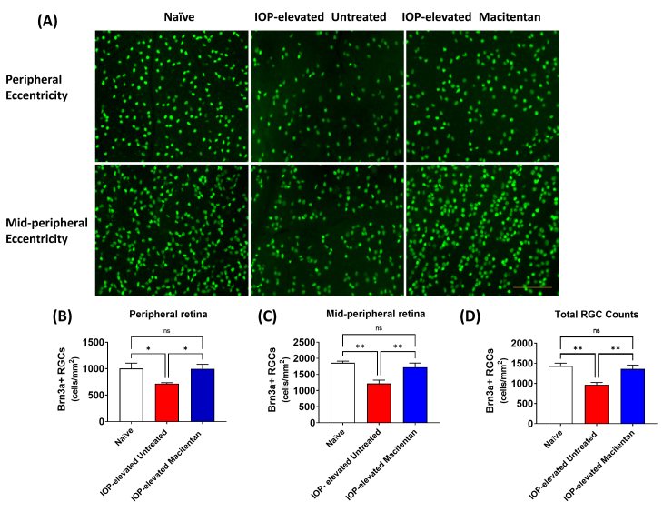
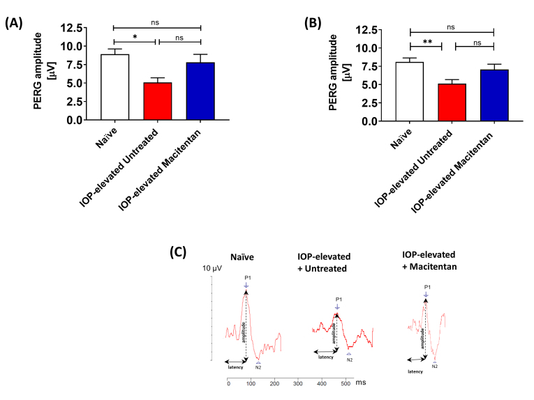
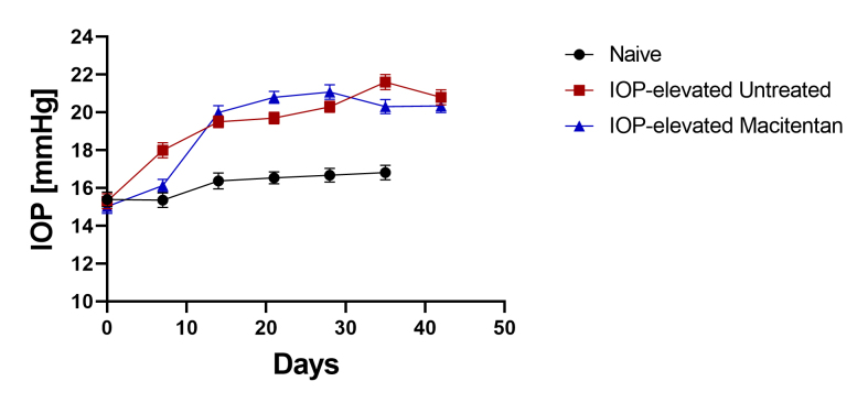
Methods: Male and female Brown Norway rats were subjected to the Morrison model of ocular hypertension by injection of hypertonic saline through the episcleral veins. Following IOP elevation, macitentan (5 mg/kg body wt) was administered orally 3 days per week, and rats with IOP elevation were maintained for 4 weeks. RGC function was determined by pattern electroretinography (PERG) at 2 and 4 weeks post-IOP elevation. Rats were euthanized by approved humane methods, and retinal flat mounts were generated and immunostained for the RGC-selective marker Brn3a. PPD-stained optic nerve sections were imaged by confocal microscopy. RGC and axon counts were conducted in a masked manner and compared between the treatment groups.
Results: Significant protection against loss of RGCs and optic nerve axons was found following oral administration of macitentan in rats with elevated IOP. In addition, a protective trend for RGC function, as measured by pattern ERG analysis, was evident following macitentan treatment.
Conclusions: Macitentan treatment had a neuroprotective effect on RGCs and their axons, independent of its IOP-lowering effect, suggesting that macitentan may complement existing treatments to prevent neurodegeneration during ocular hypertension. The findings presented have implications for the use of macitentan as an oral formulation to promote neuroprotection in glaucoma patients.
期刊介绍:
Molecular Vision is a peer-reviewed journal dedicated to the dissemination of research results in molecular biology, cell biology, and the genetics of the visual system (ocular and cortical).
Molecular Vision publishes articles presenting original research that has not previously been published and comprehensive articles reviewing the current status of a particular field or topic. Submissions to Molecular Vision are subjected to rigorous peer review. Molecular Vision does NOT publish preprints.
For authors, Molecular Vision provides a rapid means of communicating important results. Access to Molecular Vision is free and unrestricted, allowing the widest possible audience for your article. Digital publishing allows you to use color images freely (and without fees). Additionally, you may publish animations, sounds, or other supplementary information that clarifies or supports your article. Each of the authors of an article may also list an electronic mail address (which will be updated upon request) to give interested readers easy access to authors.
 求助内容:
求助内容: 应助结果提醒方式:


