Emmanuel K M Edzie, Klenam Dzefi-Tettey, Philip N Gorleku, Edmund K Brakohiapa, Peter Appiah-Thompson, Kwasi Agyen-Mensah, Michael K Amedi, Frank Quarshie, Evans Boadi, Joshua M Kpobi, Richard A Edzie, Abdul R Asemah
{"title":"加纳第三医院颅内梗死的计算机断层扫描模式。","authors":"Emmanuel K M Edzie, Klenam Dzefi-Tettey, Philip N Gorleku, Edmund K Brakohiapa, Peter Appiah-Thompson, Kwasi Agyen-Mensah, Michael K Amedi, Frank Quarshie, Evans Boadi, Joshua M Kpobi, Richard A Edzie, Abdul R Asemah","doi":"10.4314/gmj.v56i1.5","DOIUrl":null,"url":null,"abstract":"<p><strong>Objective: </strong>To determine the Computed Tomography (CT) patterns of intracranial infarcts.</p><p><strong>Design: </strong>A retrospective cross-sectional study.</p><p><strong>Setting: </strong>The CT scan unit of the Radiology Department, Cape Coast Teaching Hospital (CCTH), from February 2017 to February 2021.</p><p><strong>Participants: </strong>One thousand, one hundred and twenty-five patients with non-contrast head CT scan diagnosis of ischaemic strokes, consecutively selected over the study period without any exclusions.</p><p><strong>Main outcome measures: </strong>Patterns of non-contrast head CT scan of ischaemic strokes.</p><p><strong>Results: </strong>About 50.6% of the study participants were females with an average age of 62.59±13.91 years. Males were affected with ischaemic strokes earlier than females (<i>p<</i>0.001). The risk factors considered were, hyperlipidaemia (59.5%), hypertension (49.0%), Type 2 diabetes mellitus (DM-2) (39.6%) and smoking (3.0%). The three commonest ischaemic stroke CT scan features were wedge-shaped hypodensity extending to the edge of the brain (62.8%), sulcal flattening/effacement (57.6%) and loss of grey-white matter differentiation (51.0%), which were all significantly associated with hypertension. Small deep brain hypodensities, the rarest feature (2.2%), had no significant association with any of the risk factors considered in the study.</p><p><strong>Conclusion: </strong>Apart from the loss of grey-white matter differentiation, there was no significant association between the other CT scan features and sex. Generally, most of the risk factors and the CT scan features were significantly associated with increasing age.</p><p><strong>Funding: </strong>None declared.</p>","PeriodicalId":35509,"journal":{"name":"Ghana Medical Journal","volume":"56 1","pages":"28-37"},"PeriodicalIF":0.0000,"publicationDate":"2022-03-01","publicationTypes":"Journal Article","fieldsOfStudy":null,"isOpenAccess":false,"openAccessPdf":"https://www.ncbi.nlm.nih.gov/pmc/articles/PMC9334949/pdf/","citationCount":"0","resultStr":"{\"title\":\"Computed tomography patterns of intracranial infarcts in a Ghanaian tertiary facility.\",\"authors\":\"Emmanuel K M Edzie, Klenam Dzefi-Tettey, Philip N Gorleku, Edmund K Brakohiapa, Peter Appiah-Thompson, Kwasi Agyen-Mensah, Michael K Amedi, Frank Quarshie, Evans Boadi, Joshua M Kpobi, Richard A Edzie, Abdul R Asemah\",\"doi\":\"10.4314/gmj.v56i1.5\",\"DOIUrl\":null,\"url\":null,\"abstract\":\"<p><strong>Objective: </strong>To determine the Computed Tomography (CT) patterns of intracranial infarcts.</p><p><strong>Design: </strong>A retrospective cross-sectional study.</p><p><strong>Setting: </strong>The CT scan unit of the Radiology Department, Cape Coast Teaching Hospital (CCTH), from February 2017 to February 2021.</p><p><strong>Participants: </strong>One thousand, one hundred and twenty-five patients with non-contrast head CT scan diagnosis of ischaemic strokes, consecutively selected over the study period without any exclusions.</p><p><strong>Main outcome measures: </strong>Patterns of non-contrast head CT scan of ischaemic strokes.</p><p><strong>Results: </strong>About 50.6% of the study participants were females with an average age of 62.59±13.91 years. Males were affected with ischaemic strokes earlier than females (<i>p<</i>0.001). The risk factors considered were, hyperlipidaemia (59.5%), hypertension (49.0%), Type 2 diabetes mellitus (DM-2) (39.6%) and smoking (3.0%). The three commonest ischaemic stroke CT scan features were wedge-shaped hypodensity extending to the edge of the brain (62.8%), sulcal flattening/effacement (57.6%) and loss of grey-white matter differentiation (51.0%), which were all significantly associated with hypertension. Small deep brain hypodensities, the rarest feature (2.2%), had no significant association with any of the risk factors considered in the study.</p><p><strong>Conclusion: </strong>Apart from the loss of grey-white matter differentiation, there was no significant association between the other CT scan features and sex. Generally, most of the risk factors and the CT scan features were significantly associated with increasing age.</p><p><strong>Funding: </strong>None declared.</p>\",\"PeriodicalId\":35509,\"journal\":{\"name\":\"Ghana Medical Journal\",\"volume\":\"56 1\",\"pages\":\"28-37\"},\"PeriodicalIF\":0.0000,\"publicationDate\":\"2022-03-01\",\"publicationTypes\":\"Journal Article\",\"fieldsOfStudy\":null,\"isOpenAccess\":false,\"openAccessPdf\":\"https://www.ncbi.nlm.nih.gov/pmc/articles/PMC9334949/pdf/\",\"citationCount\":\"0\",\"resultStr\":null,\"platform\":\"Semanticscholar\",\"paperid\":null,\"PeriodicalName\":\"Ghana Medical Journal\",\"FirstCategoryId\":\"1085\",\"ListUrlMain\":\"https://doi.org/10.4314/gmj.v56i1.5\",\"RegionNum\":0,\"RegionCategory\":null,\"ArticlePicture\":[],\"TitleCN\":null,\"AbstractTextCN\":null,\"PMCID\":null,\"EPubDate\":\"\",\"PubModel\":\"\",\"JCR\":\"Q3\",\"JCRName\":\"Medicine\",\"Score\":null,\"Total\":0}","platform":"Semanticscholar","paperid":null,"PeriodicalName":"Ghana Medical Journal","FirstCategoryId":"1085","ListUrlMain":"https://doi.org/10.4314/gmj.v56i1.5","RegionNum":0,"RegionCategory":null,"ArticlePicture":[],"TitleCN":null,"AbstractTextCN":null,"PMCID":null,"EPubDate":"","PubModel":"","JCR":"Q3","JCRName":"Medicine","Score":null,"Total":0}
Computed tomography patterns of intracranial infarcts in a Ghanaian tertiary facility.
Objective: To determine the Computed Tomography (CT) patterns of intracranial infarcts.
Design: A retrospective cross-sectional study.
Setting: The CT scan unit of the Radiology Department, Cape Coast Teaching Hospital (CCTH), from February 2017 to February 2021.
Participants: One thousand, one hundred and twenty-five patients with non-contrast head CT scan diagnosis of ischaemic strokes, consecutively selected over the study period without any exclusions.
Main outcome measures: Patterns of non-contrast head CT scan of ischaemic strokes.
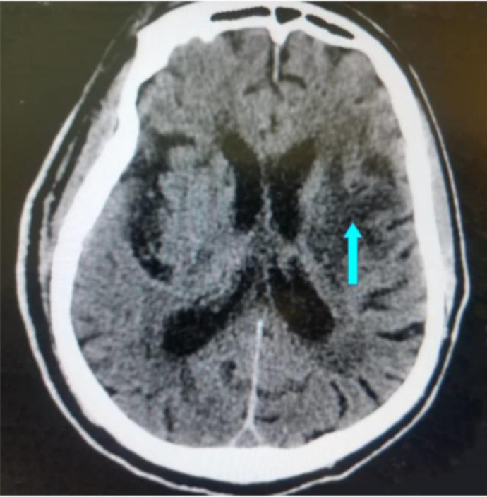
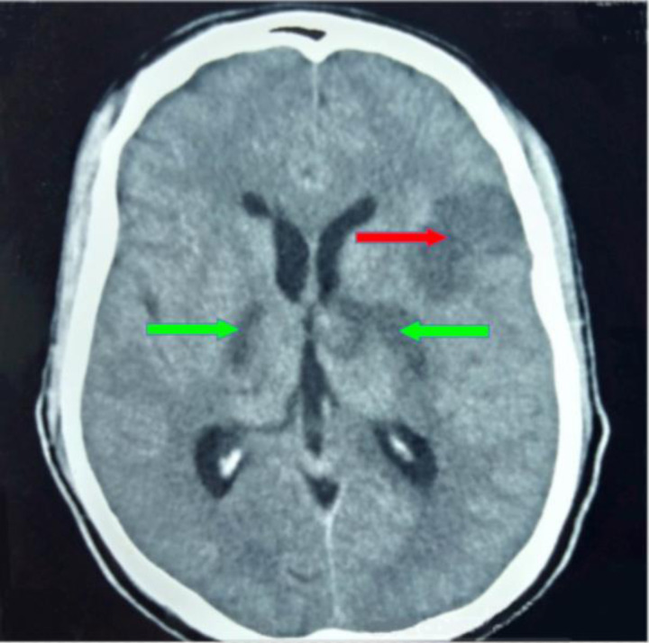
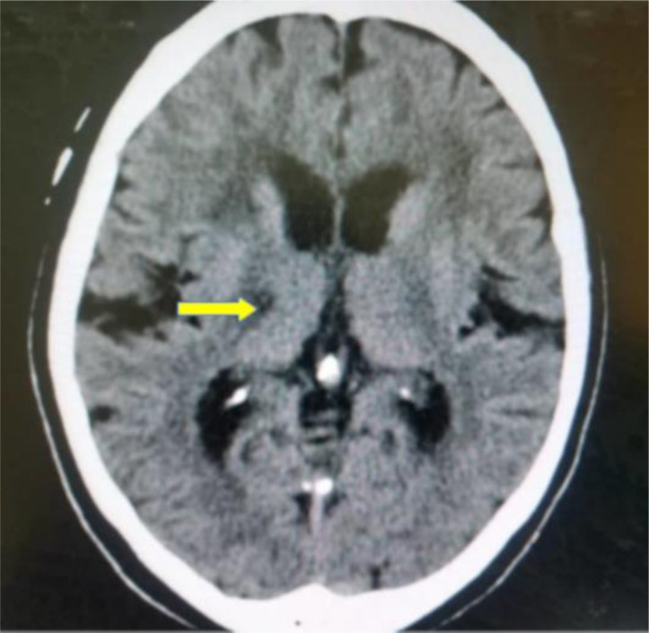
Results: About 50.6% of the study participants were females with an average age of 62.59±13.91 years. Males were affected with ischaemic strokes earlier than females (p<0.001). The risk factors considered were, hyperlipidaemia (59.5%), hypertension (49.0%), Type 2 diabetes mellitus (DM-2) (39.6%) and smoking (3.0%). The three commonest ischaemic stroke CT scan features were wedge-shaped hypodensity extending to the edge of the brain (62.8%), sulcal flattening/effacement (57.6%) and loss of grey-white matter differentiation (51.0%), which were all significantly associated with hypertension. Small deep brain hypodensities, the rarest feature (2.2%), had no significant association with any of the risk factors considered in the study.
Conclusion: Apart from the loss of grey-white matter differentiation, there was no significant association between the other CT scan features and sex. Generally, most of the risk factors and the CT scan features were significantly associated with increasing age.

 求助内容:
求助内容: 应助结果提醒方式:


