Deepaysh D C S Dutt, Seyhan Yazar, Jason Charng, David A Mackey, Fred K Chen, Danuta M Sampson
{"title":"校正光学相干断层扫描血管造影图像的中央凹无血管区面积测量的放大误差与估计轴向长度。","authors":"Deepaysh D C S Dutt, Seyhan Yazar, Jason Charng, David A Mackey, Fred K Chen, Danuta M Sampson","doi":"10.1186/s40662-022-00299-x","DOIUrl":null,"url":null,"abstract":"<p><strong>Background: </strong>To generate and validate a method to estimate axial length estimated (AL<sub>est</sub>) from spherical equivalent (SE) and corneal curvature [keratometry (K)], and to determine if this AL<sub>est</sub> can replace actual axial length (AL<sub>act</sub>) for correcting transverse magnification error in optical coherence tomography angiography (OCTA) images using the Littmann-Bennett formula.</p><p><strong>Methods: </strong>Data from 1301 participants of the Raine Study Gen2-20 year follow-up were divided into two datasets to generate (n = 650) and validate (n = 651) a relationship between AL, SE, and K. The developed formula was then applied to a separate dataset of 46 participants with AL, SE, and K measurements and OCTA images to estimate and compare the performance of AL<sub>est</sub> against AL<sub>act</sub> in correcting transverse magnification error in OCTA images when measuring the foveal avascular zone area (FAZA).</p><p><strong>Results: </strong>The formula for AL<sub>est</sub> yielded the equation: AL<sub>est</sub> = 2.102K - 0.4125SE + 7.268, R<sup>2</sup> = 0.794. There was good agreement between AL<sub>est</sub> and AL<sub>act</sub> for both study cohorts. The mean difference [standard deviation (SD)] between FAZA corrected with AL<sub>est</sub> and AL<sub>act</sub> was 0.002 (0.015) mm<sup>2</sup> with the 95% limits of agreement (LoA) of - 0.027 to 0.031 mm<sup>2</sup>. In comparison, mean difference (SD) between FAZA uncorrected and corrected with AL<sub>act</sub> was - 0.005 (0.030) mm<sup>2</sup>, with 95% LoA of - 0.064 to 0.054 mm<sup>2</sup>.</p><p><strong>Conclusions: </strong>AL<sub>act</sub> is more accurate than AL<sub>est</sub> and hence should be used preferentially in magnification error correction in the clinical setting. FAZA corrected with AL<sub>est</sub> is comparable to FAZA corrected with AL<sub>act</sub>, while FAZA measurements using images corrected with AL<sub>est</sub> have a greater accuracy than measurements on uncorrected images. Hence, in the absence of AL<sub>act</sub>, clinicians should use AL<sub>est</sub> to correct for magnification error as this provides for more accurate measurements of fundus parameters than uncorrected images.</p>","PeriodicalId":520624,"journal":{"name":"Eye and vision (London, England)","volume":" ","pages":"29"},"PeriodicalIF":0.0000,"publicationDate":"2022-08-01","publicationTypes":"Journal Article","fieldsOfStudy":null,"isOpenAccess":false,"openAccessPdf":"https://www.ncbi.nlm.nih.gov/pmc/articles/PMC9341098/pdf/","citationCount":"0","resultStr":"{\"title\":\"Correcting magnification error in foveal avascular zone area measurements of optical coherence tomography angiography images with estimated axial length.\",\"authors\":\"Deepaysh D C S Dutt, Seyhan Yazar, Jason Charng, David A Mackey, Fred K Chen, Danuta M Sampson\",\"doi\":\"10.1186/s40662-022-00299-x\",\"DOIUrl\":null,\"url\":null,\"abstract\":\"<p><strong>Background: </strong>To generate and validate a method to estimate axial length estimated (AL<sub>est</sub>) from spherical equivalent (SE) and corneal curvature [keratometry (K)], and to determine if this AL<sub>est</sub> can replace actual axial length (AL<sub>act</sub>) for correcting transverse magnification error in optical coherence tomography angiography (OCTA) images using the Littmann-Bennett formula.</p><p><strong>Methods: </strong>Data from 1301 participants of the Raine Study Gen2-20 year follow-up were divided into two datasets to generate (n = 650) and validate (n = 651) a relationship between AL, SE, and K. The developed formula was then applied to a separate dataset of 46 participants with AL, SE, and K measurements and OCTA images to estimate and compare the performance of AL<sub>est</sub> against AL<sub>act</sub> in correcting transverse magnification error in OCTA images when measuring the foveal avascular zone area (FAZA).</p><p><strong>Results: </strong>The formula for AL<sub>est</sub> yielded the equation: AL<sub>est</sub> = 2.102K - 0.4125SE + 7.268, R<sup>2</sup> = 0.794. There was good agreement between AL<sub>est</sub> and AL<sub>act</sub> for both study cohorts. The mean difference [standard deviation (SD)] between FAZA corrected with AL<sub>est</sub> and AL<sub>act</sub> was 0.002 (0.015) mm<sup>2</sup> with the 95% limits of agreement (LoA) of - 0.027 to 0.031 mm<sup>2</sup>. In comparison, mean difference (SD) between FAZA uncorrected and corrected with AL<sub>act</sub> was - 0.005 (0.030) mm<sup>2</sup>, with 95% LoA of - 0.064 to 0.054 mm<sup>2</sup>.</p><p><strong>Conclusions: </strong>AL<sub>act</sub> is more accurate than AL<sub>est</sub> and hence should be used preferentially in magnification error correction in the clinical setting. FAZA corrected with AL<sub>est</sub> is comparable to FAZA corrected with AL<sub>act</sub>, while FAZA measurements using images corrected with AL<sub>est</sub> have a greater accuracy than measurements on uncorrected images. Hence, in the absence of AL<sub>act</sub>, clinicians should use AL<sub>est</sub> to correct for magnification error as this provides for more accurate measurements of fundus parameters than uncorrected images.</p>\",\"PeriodicalId\":520624,\"journal\":{\"name\":\"Eye and vision (London, England)\",\"volume\":\" \",\"pages\":\"29\"},\"PeriodicalIF\":0.0000,\"publicationDate\":\"2022-08-01\",\"publicationTypes\":\"Journal Article\",\"fieldsOfStudy\":null,\"isOpenAccess\":false,\"openAccessPdf\":\"https://www.ncbi.nlm.nih.gov/pmc/articles/PMC9341098/pdf/\",\"citationCount\":\"0\",\"resultStr\":null,\"platform\":\"Semanticscholar\",\"paperid\":null,\"PeriodicalName\":\"Eye and vision (London, England)\",\"FirstCategoryId\":\"3\",\"ListUrlMain\":\"https://doi.org/10.1186/s40662-022-00299-x\",\"RegionNum\":0,\"RegionCategory\":null,\"ArticlePicture\":[],\"TitleCN\":null,\"AbstractTextCN\":null,\"PMCID\":null,\"EPubDate\":\"\",\"PubModel\":\"\",\"JCR\":\"\",\"JCRName\":\"\",\"Score\":null,\"Total\":0}","platform":"Semanticscholar","paperid":null,"PeriodicalName":"Eye and vision (London, England)","FirstCategoryId":"3","ListUrlMain":"https://doi.org/10.1186/s40662-022-00299-x","RegionNum":0,"RegionCategory":null,"ArticlePicture":[],"TitleCN":null,"AbstractTextCN":null,"PMCID":null,"EPubDate":"","PubModel":"","JCR":"","JCRName":"","Score":null,"Total":0}
Correcting magnification error in foveal avascular zone area measurements of optical coherence tomography angiography images with estimated axial length.
Background: To generate and validate a method to estimate axial length estimated (ALest) from spherical equivalent (SE) and corneal curvature [keratometry (K)], and to determine if this ALest can replace actual axial length (ALact) for correcting transverse magnification error in optical coherence tomography angiography (OCTA) images using the Littmann-Bennett formula.
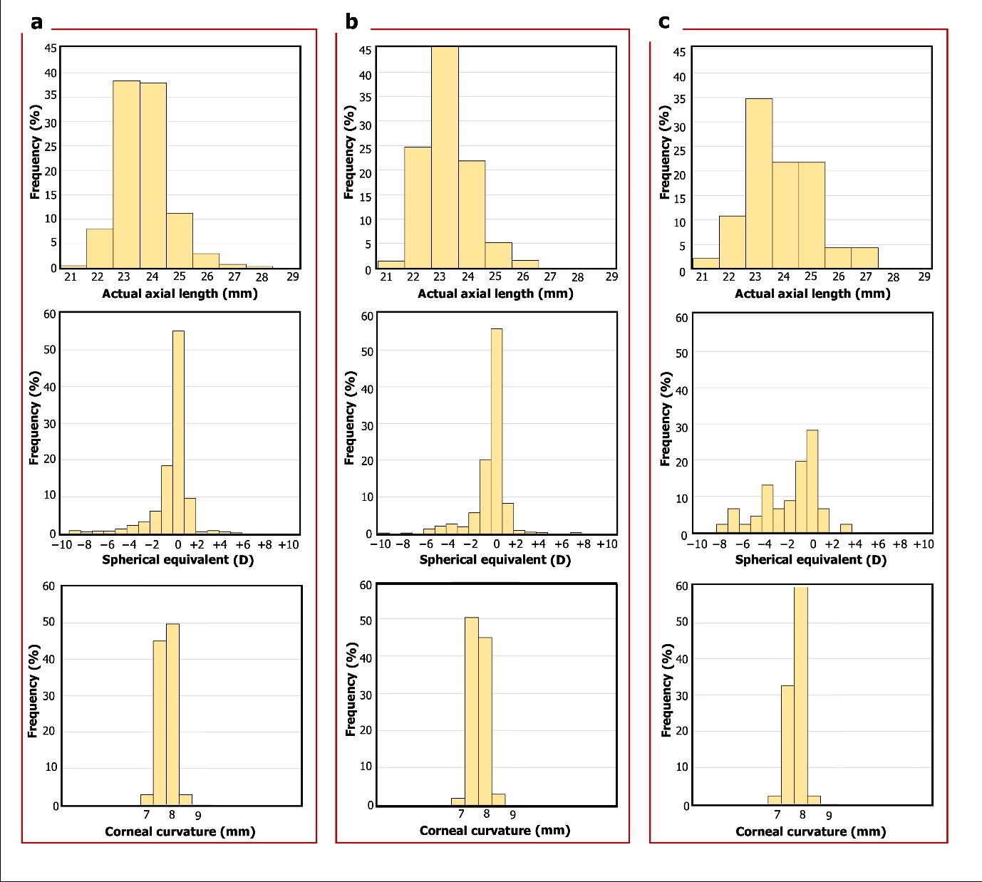
Methods: Data from 1301 participants of the Raine Study Gen2-20 year follow-up were divided into two datasets to generate (n = 650) and validate (n = 651) a relationship between AL, SE, and K. The developed formula was then applied to a separate dataset of 46 participants with AL, SE, and K measurements and OCTA images to estimate and compare the performance of ALest against ALact in correcting transverse magnification error in OCTA images when measuring the foveal avascular zone area (FAZA).
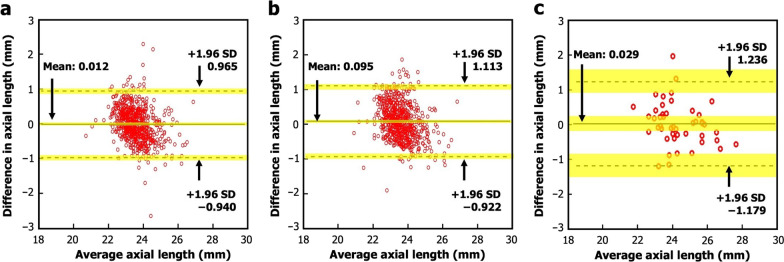
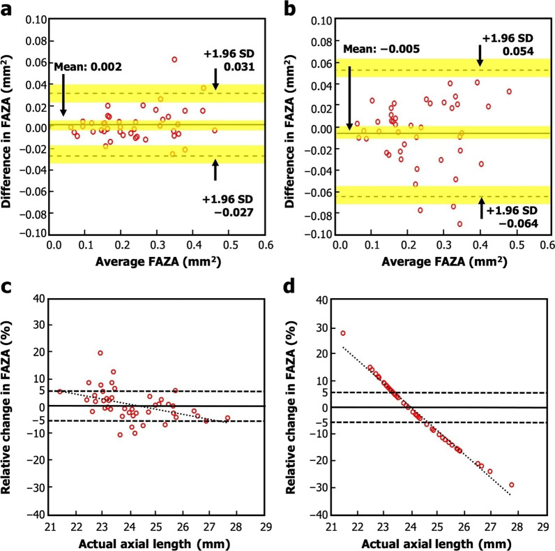
Results: The formula for ALest yielded the equation: ALest = 2.102K - 0.4125SE + 7.268, R2 = 0.794. There was good agreement between ALest and ALact for both study cohorts. The mean difference [standard deviation (SD)] between FAZA corrected with ALest and ALact was 0.002 (0.015) mm2 with the 95% limits of agreement (LoA) of - 0.027 to 0.031 mm2. In comparison, mean difference (SD) between FAZA uncorrected and corrected with ALact was - 0.005 (0.030) mm2, with 95% LoA of - 0.064 to 0.054 mm2.
Conclusions: ALact is more accurate than ALest and hence should be used preferentially in magnification error correction in the clinical setting. FAZA corrected with ALest is comparable to FAZA corrected with ALact, while FAZA measurements using images corrected with ALest have a greater accuracy than measurements on uncorrected images. Hence, in the absence of ALact, clinicians should use ALest to correct for magnification error as this provides for more accurate measurements of fundus parameters than uncorrected images.

 求助内容:
求助内容: 应助结果提醒方式:


