Jihong Zhou, Wei Gu, Yan Gao, Wenjuan Wang, Fengju Zhang
{"title":"低至中度近视小切口晶状体摘除术及飞秒激光辅助激光原位角膜移植术后近视消退的生存分析。","authors":"Jihong Zhou, Wei Gu, Yan Gao, Wenjuan Wang, Fengju Zhang","doi":"10.1186/s40662-022-00300-7","DOIUrl":null,"url":null,"abstract":"<p><strong>Background: </strong>To report the predictive factors of myopic regression in patients who had undergone small incision lenticular extraction (SMILE) and femtosecond laser-assisted laser in situ keratomileuses (FS-LASIK) after 3-12 months of follow-up.</p><p><strong>Methods: </strong>This retrospective case series study recruited patients with a subjective sphere of - 1.00 to - 6.00 D myopia. SMILE was performed in 1629 eyes of 1629 patients with a subjective refraction spherical equivalent (SEQ) of - 4.57 ± 1.20 D and 1414 eyes of 1414 patients with a subjective SEQ of - 4.53 ± 1.26 D in FS-LASIK. Refractive outcomes were recorded at 1 day, 1 week, and 1, 3, 6, and 12 months postoperatively. Predictors affecting myopic regression and other covariates were estimated with a Cox proportional hazard (Cox PH) model for the two surgical methods.</p><p><strong>Results: </strong>At 12 months, no significant difference was evident in the efficacy (P = 0.934), predictability (P = 0.733), or stability (P = 0.66) between FS-LASIK and SMILE. The survival rates were 83.7% in the FS-LASIK group and 88.1% in the SMILE group. Multivariate analysis by the Cox PH model revealed a similar probability of postoperative myopic regression with SMILE or FS-LASIK (P = 0.630). Predictors of myopic regression included preoperative higher-order aberration root mean square with 3 mm pupil diameter (pre-HOA-RMS<sub>3</sub>) (P = 0.004), anterior chamber depth (ACD) (P = 0.015), pre-subjective sphere (P = 0.016), corneal diameter (P = 0.016), optical zone (OZ) (P = 0.02), and predicted depth of ablation (DA) (P = 0.003).</p><p><strong>Conclusion: </strong>SMILE and FS-LASIK had a similar risk of myopic regression for low to moderate myopia. Pre-HOA-RMS<sub>3</sub>, ACD, pre-subjective sphere, corneal diameter, OZ, and predicted DA were predictors of myopic regression.</p>","PeriodicalId":520624,"journal":{"name":"Eye and vision (London, England)","volume":" ","pages":"28"},"PeriodicalIF":0.0000,"publicationDate":"2022-08-01","publicationTypes":"Journal Article","fieldsOfStudy":null,"isOpenAccess":false,"openAccessPdf":"https://www.ncbi.nlm.nih.gov/pmc/articles/PMC9341088/pdf/","citationCount":"0","resultStr":"{\"title\":\"Survival analysis of myopic regression after small incision lenticule extraction and femtosecond laser-assisted laser in situ keratomileusis for low to moderate myopia.\",\"authors\":\"Jihong Zhou, Wei Gu, Yan Gao, Wenjuan Wang, Fengju Zhang\",\"doi\":\"10.1186/s40662-022-00300-7\",\"DOIUrl\":null,\"url\":null,\"abstract\":\"<p><strong>Background: </strong>To report the predictive factors of myopic regression in patients who had undergone small incision lenticular extraction (SMILE) and femtosecond laser-assisted laser in situ keratomileuses (FS-LASIK) after 3-12 months of follow-up.</p><p><strong>Methods: </strong>This retrospective case series study recruited patients with a subjective sphere of - 1.00 to - 6.00 D myopia. SMILE was performed in 1629 eyes of 1629 patients with a subjective refraction spherical equivalent (SEQ) of - 4.57 ± 1.20 D and 1414 eyes of 1414 patients with a subjective SEQ of - 4.53 ± 1.26 D in FS-LASIK. Refractive outcomes were recorded at 1 day, 1 week, and 1, 3, 6, and 12 months postoperatively. Predictors affecting myopic regression and other covariates were estimated with a Cox proportional hazard (Cox PH) model for the two surgical methods.</p><p><strong>Results: </strong>At 12 months, no significant difference was evident in the efficacy (P = 0.934), predictability (P = 0.733), or stability (P = 0.66) between FS-LASIK and SMILE. The survival rates were 83.7% in the FS-LASIK group and 88.1% in the SMILE group. Multivariate analysis by the Cox PH model revealed a similar probability of postoperative myopic regression with SMILE or FS-LASIK (P = 0.630). Predictors of myopic regression included preoperative higher-order aberration root mean square with 3 mm pupil diameter (pre-HOA-RMS<sub>3</sub>) (P = 0.004), anterior chamber depth (ACD) (P = 0.015), pre-subjective sphere (P = 0.016), corneal diameter (P = 0.016), optical zone (OZ) (P = 0.02), and predicted depth of ablation (DA) (P = 0.003).</p><p><strong>Conclusion: </strong>SMILE and FS-LASIK had a similar risk of myopic regression for low to moderate myopia. Pre-HOA-RMS<sub>3</sub>, ACD, pre-subjective sphere, corneal diameter, OZ, and predicted DA were predictors of myopic regression.</p>\",\"PeriodicalId\":520624,\"journal\":{\"name\":\"Eye and vision (London, England)\",\"volume\":\" \",\"pages\":\"28\"},\"PeriodicalIF\":0.0000,\"publicationDate\":\"2022-08-01\",\"publicationTypes\":\"Journal Article\",\"fieldsOfStudy\":null,\"isOpenAccess\":false,\"openAccessPdf\":\"https://www.ncbi.nlm.nih.gov/pmc/articles/PMC9341088/pdf/\",\"citationCount\":\"0\",\"resultStr\":null,\"platform\":\"Semanticscholar\",\"paperid\":null,\"PeriodicalName\":\"Eye and vision (London, England)\",\"FirstCategoryId\":\"3\",\"ListUrlMain\":\"https://doi.org/10.1186/s40662-022-00300-7\",\"RegionNum\":0,\"RegionCategory\":null,\"ArticlePicture\":[],\"TitleCN\":null,\"AbstractTextCN\":null,\"PMCID\":null,\"EPubDate\":\"\",\"PubModel\":\"\",\"JCR\":\"\",\"JCRName\":\"\",\"Score\":null,\"Total\":0}","platform":"Semanticscholar","paperid":null,"PeriodicalName":"Eye and vision (London, England)","FirstCategoryId":"3","ListUrlMain":"https://doi.org/10.1186/s40662-022-00300-7","RegionNum":0,"RegionCategory":null,"ArticlePicture":[],"TitleCN":null,"AbstractTextCN":null,"PMCID":null,"EPubDate":"","PubModel":"","JCR":"","JCRName":"","Score":null,"Total":0}
Survival analysis of myopic regression after small incision lenticule extraction and femtosecond laser-assisted laser in situ keratomileusis for low to moderate myopia.
Background: To report the predictive factors of myopic regression in patients who had undergone small incision lenticular extraction (SMILE) and femtosecond laser-assisted laser in situ keratomileuses (FS-LASIK) after 3-12 months of follow-up.
Methods: This retrospective case series study recruited patients with a subjective sphere of - 1.00 to - 6.00 D myopia. SMILE was performed in 1629 eyes of 1629 patients with a subjective refraction spherical equivalent (SEQ) of - 4.57 ± 1.20 D and 1414 eyes of 1414 patients with a subjective SEQ of - 4.53 ± 1.26 D in FS-LASIK. Refractive outcomes were recorded at 1 day, 1 week, and 1, 3, 6, and 12 months postoperatively. Predictors affecting myopic regression and other covariates were estimated with a Cox proportional hazard (Cox PH) model for the two surgical methods.
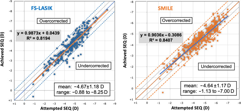
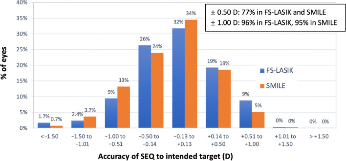
Results: At 12 months, no significant difference was evident in the efficacy (P = 0.934), predictability (P = 0.733), or stability (P = 0.66) between FS-LASIK and SMILE. The survival rates were 83.7% in the FS-LASIK group and 88.1% in the SMILE group. Multivariate analysis by the Cox PH model revealed a similar probability of postoperative myopic regression with SMILE or FS-LASIK (P = 0.630). Predictors of myopic regression included preoperative higher-order aberration root mean square with 3 mm pupil diameter (pre-HOA-RMS3) (P = 0.004), anterior chamber depth (ACD) (P = 0.015), pre-subjective sphere (P = 0.016), corneal diameter (P = 0.016), optical zone (OZ) (P = 0.02), and predicted depth of ablation (DA) (P = 0.003).
Conclusion: SMILE and FS-LASIK had a similar risk of myopic regression for low to moderate myopia. Pre-HOA-RMS3, ACD, pre-subjective sphere, corneal diameter, OZ, and predicted DA were predictors of myopic regression.


 求助内容:
求助内容: 应助结果提醒方式:


