Jérémy Morille, Marion Mandon, Stéphane Rodriguez, David Roulois, Simon Leonard, Alexandra Garcia, Sandrine Wiertlewski, Emmanuelle Le Page, Laureline Berthelot, Arnaud Nicot, Camille Mathé, Flora Lejeune, Karin Tarte, Céline Delaloy, Patricia Amé, David Laplaud, Laure Michel
{"title":"多发性硬化症CSF富含滤泡T细胞,显示Th1/Eomes特征。","authors":"Jérémy Morille, Marion Mandon, Stéphane Rodriguez, David Roulois, Simon Leonard, Alexandra Garcia, Sandrine Wiertlewski, Emmanuelle Le Page, Laureline Berthelot, Arnaud Nicot, Camille Mathé, Flora Lejeune, Karin Tarte, Céline Delaloy, Patricia Amé, David Laplaud, Laure Michel","doi":"10.1212/NXI.0000000000200033","DOIUrl":null,"url":null,"abstract":"<p><strong>Background and objectives: </strong>Tertiary lymphoid structures and aggregates are reported in the meninges of patients with multiple sclerosis (MS), especially at the progressive stage, and are strongly associated with cortical lesions and disability. Besides B cells, these structures comprise follicular helper T (Tfh) cells that are crucial to support B-cell differentiation. Tfh cells play a pivotal role in amplifying autoreactive B cells and promoting autoantibody production in several autoimmune diseases, but very few are known in MS. In this study, we examined the phenotype, frequency, and transcriptome of circulating cTfh cells in the blood and CSF of patients with relapsing-remitting MS (RRMS).</p><p><strong>Methods: </strong>The phenotype and frequency of cTfh cells were analyzed in the blood of 39 healthy controls and 41 untreated patients with RRMS and in the CSF and paired blood of 10 patients with drug-naive RRMS at diagnosis by flow cytometry. Using an in vitro model of blood-brain barrier, we assessed the transendothelial migratory abilities of the different cTfh-cell subsets. Finally, we performed an RNA sequencing analysis of paired CSF cTfh cells and blood cTfh cells in 8 patients sampled at their first demyelinating event.</p><p><strong>Results: </strong>The blood phenotype and frequency of cTfh cells were not significantly modified in patients with RRMS. In the CSF, we found an important infiltration of Tfh1 cells, with a high proportion of activated PD1<sup>+</sup> cells. We demonstrated that the specific subset of Tfh1 cells presents increased migration abilities to cross an in vitro model of blood-brain barrier. Of interest, even at the first demyelinating event, cTfh cells in the CSF display specific characteristics with upregulation of <i>EOMES</i> gene and proinflammatory/cytotoxic transcriptomic signature able to efficiently distinguish cTfh cells from the CSF and blood. Finally, interactome analysis revealed potential strong cross talk between pathogenic B cells and CSF cTfh cells, pointing out the CSF as opportune supportive compartment and highlighting the very early implication of B-cell helper T cells in MS pathogenesis.</p><p><strong>Discussion: </strong>Overall, CSF enrichment in activated Tfh1 as soon as disease diagnosis, associated with high expression of EOMES, and a predicted high propensity to interact with CSF B cells suggest that these cells probably contribute to disease onset and/or activity.</p>","PeriodicalId":520720,"journal":{"name":"Neurology(R) neuroimmunology & neuroinflammation","volume":" ","pages":""},"PeriodicalIF":0.0000,"publicationDate":"2022-10-20","publicationTypes":"Journal Article","fieldsOfStudy":null,"isOpenAccess":false,"openAccessPdf":"https://ftp.ncbi.nlm.nih.gov/pub/pmc/oa_pdf/47/3a/NXI-2022-200039.PMC9585484.pdf","citationCount":"5","resultStr":"{\"title\":\"Multiple Sclerosis CSF Is Enriched With Follicular T Cells Displaying a Th1/Eomes Signature.\",\"authors\":\"Jérémy Morille, Marion Mandon, Stéphane Rodriguez, David Roulois, Simon Leonard, Alexandra Garcia, Sandrine Wiertlewski, Emmanuelle Le Page, Laureline Berthelot, Arnaud Nicot, Camille Mathé, Flora Lejeune, Karin Tarte, Céline Delaloy, Patricia Amé, David Laplaud, Laure Michel\",\"doi\":\"10.1212/NXI.0000000000200033\",\"DOIUrl\":null,\"url\":null,\"abstract\":\"<p><strong>Background and objectives: </strong>Tertiary lymphoid structures and aggregates are reported in the meninges of patients with multiple sclerosis (MS), especially at the progressive stage, and are strongly associated with cortical lesions and disability. Besides B cells, these structures comprise follicular helper T (Tfh) cells that are crucial to support B-cell differentiation. Tfh cells play a pivotal role in amplifying autoreactive B cells and promoting autoantibody production in several autoimmune diseases, but very few are known in MS. In this study, we examined the phenotype, frequency, and transcriptome of circulating cTfh cells in the blood and CSF of patients with relapsing-remitting MS (RRMS).</p><p><strong>Methods: </strong>The phenotype and frequency of cTfh cells were analyzed in the blood of 39 healthy controls and 41 untreated patients with RRMS and in the CSF and paired blood of 10 patients with drug-naive RRMS at diagnosis by flow cytometry. Using an in vitro model of blood-brain barrier, we assessed the transendothelial migratory abilities of the different cTfh-cell subsets. Finally, we performed an RNA sequencing analysis of paired CSF cTfh cells and blood cTfh cells in 8 patients sampled at their first demyelinating event.</p><p><strong>Results: </strong>The blood phenotype and frequency of cTfh cells were not significantly modified in patients with RRMS. In the CSF, we found an important infiltration of Tfh1 cells, with a high proportion of activated PD1<sup>+</sup> cells. We demonstrated that the specific subset of Tfh1 cells presents increased migration abilities to cross an in vitro model of blood-brain barrier. Of interest, even at the first demyelinating event, cTfh cells in the CSF display specific characteristics with upregulation of <i>EOMES</i> gene and proinflammatory/cytotoxic transcriptomic signature able to efficiently distinguish cTfh cells from the CSF and blood. Finally, interactome analysis revealed potential strong cross talk between pathogenic B cells and CSF cTfh cells, pointing out the CSF as opportune supportive compartment and highlighting the very early implication of B-cell helper T cells in MS pathogenesis.</p><p><strong>Discussion: </strong>Overall, CSF enrichment in activated Tfh1 as soon as disease diagnosis, associated with high expression of EOMES, and a predicted high propensity to interact with CSF B cells suggest that these cells probably contribute to disease onset and/or activity.</p>\",\"PeriodicalId\":520720,\"journal\":{\"name\":\"Neurology(R) neuroimmunology & neuroinflammation\",\"volume\":\" \",\"pages\":\"\"},\"PeriodicalIF\":0.0000,\"publicationDate\":\"2022-10-20\",\"publicationTypes\":\"Journal Article\",\"fieldsOfStudy\":null,\"isOpenAccess\":false,\"openAccessPdf\":\"https://ftp.ncbi.nlm.nih.gov/pub/pmc/oa_pdf/47/3a/NXI-2022-200039.PMC9585484.pdf\",\"citationCount\":\"5\",\"resultStr\":null,\"platform\":\"Semanticscholar\",\"paperid\":null,\"PeriodicalName\":\"Neurology(R) neuroimmunology & neuroinflammation\",\"FirstCategoryId\":\"3\",\"ListUrlMain\":\"https://doi.org/10.1212/NXI.0000000000200033\",\"RegionNum\":0,\"RegionCategory\":null,\"ArticlePicture\":[],\"TitleCN\":null,\"AbstractTextCN\":null,\"PMCID\":null,\"EPubDate\":\"2022/11/1 0:00:00\",\"PubModel\":\"Print\",\"JCR\":\"\",\"JCRName\":\"\",\"Score\":null,\"Total\":0}","platform":"Semanticscholar","paperid":null,"PeriodicalName":"Neurology(R) neuroimmunology & neuroinflammation","FirstCategoryId":"3","ListUrlMain":"https://doi.org/10.1212/NXI.0000000000200033","RegionNum":0,"RegionCategory":null,"ArticlePicture":[],"TitleCN":null,"AbstractTextCN":null,"PMCID":null,"EPubDate":"2022/11/1 0:00:00","PubModel":"Print","JCR":"","JCRName":"","Score":null,"Total":0}
Multiple Sclerosis CSF Is Enriched With Follicular T Cells Displaying a Th1/Eomes Signature.
Background and objectives: Tertiary lymphoid structures and aggregates are reported in the meninges of patients with multiple sclerosis (MS), especially at the progressive stage, and are strongly associated with cortical lesions and disability. Besides B cells, these structures comprise follicular helper T (Tfh) cells that are crucial to support B-cell differentiation. Tfh cells play a pivotal role in amplifying autoreactive B cells and promoting autoantibody production in several autoimmune diseases, but very few are known in MS. In this study, we examined the phenotype, frequency, and transcriptome of circulating cTfh cells in the blood and CSF of patients with relapsing-remitting MS (RRMS).
Methods: The phenotype and frequency of cTfh cells were analyzed in the blood of 39 healthy controls and 41 untreated patients with RRMS and in the CSF and paired blood of 10 patients with drug-naive RRMS at diagnosis by flow cytometry. Using an in vitro model of blood-brain barrier, we assessed the transendothelial migratory abilities of the different cTfh-cell subsets. Finally, we performed an RNA sequencing analysis of paired CSF cTfh cells and blood cTfh cells in 8 patients sampled at their first demyelinating event.
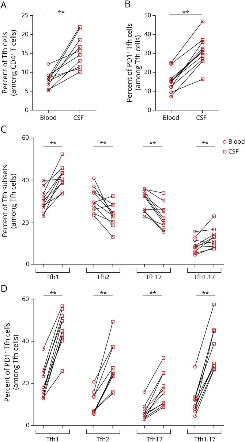
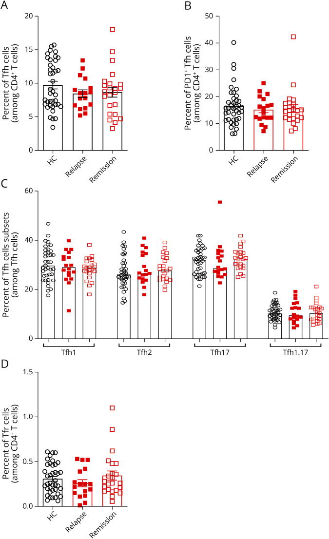
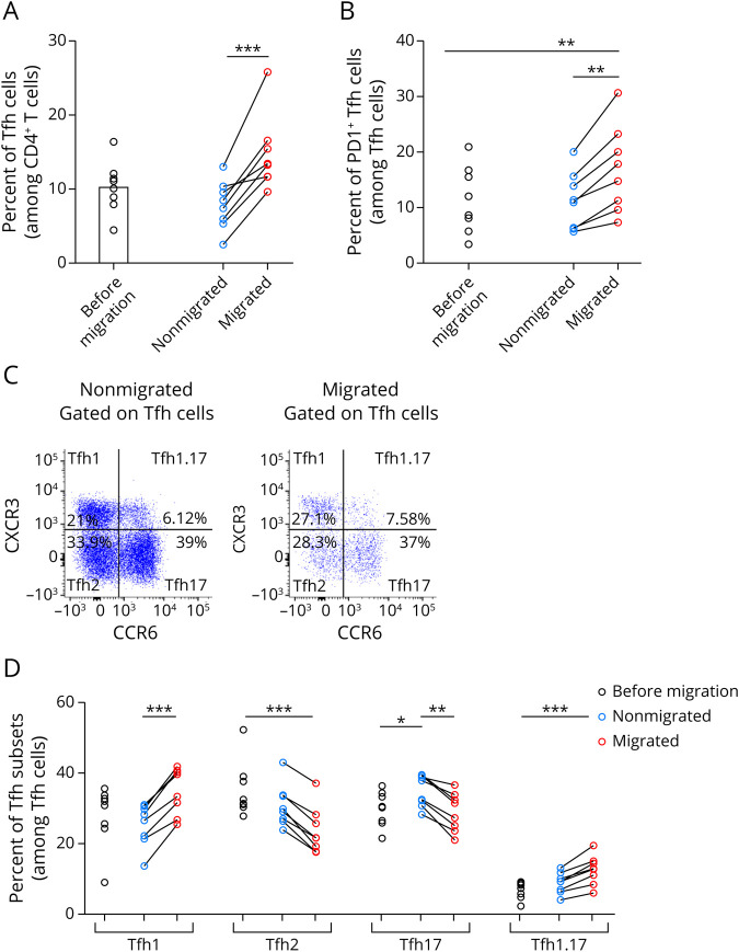
Results: The blood phenotype and frequency of cTfh cells were not significantly modified in patients with RRMS. In the CSF, we found an important infiltration of Tfh1 cells, with a high proportion of activated PD1+ cells. We demonstrated that the specific subset of Tfh1 cells presents increased migration abilities to cross an in vitro model of blood-brain barrier. Of interest, even at the first demyelinating event, cTfh cells in the CSF display specific characteristics with upregulation of EOMES gene and proinflammatory/cytotoxic transcriptomic signature able to efficiently distinguish cTfh cells from the CSF and blood. Finally, interactome analysis revealed potential strong cross talk between pathogenic B cells and CSF cTfh cells, pointing out the CSF as opportune supportive compartment and highlighting the very early implication of B-cell helper T cells in MS pathogenesis.
Discussion: Overall, CSF enrichment in activated Tfh1 as soon as disease diagnosis, associated with high expression of EOMES, and a predicted high propensity to interact with CSF B cells suggest that these cells probably contribute to disease onset and/or activity.

 求助内容:
求助内容: 应助结果提醒方式:


