Masakatsu Nohara, Mayo Iwasaki, Mahiro Nishio, Akihiko Sugiyama
{"title":"早期胚胎期卵暴露于甲氨蝶呤对日本鹌鹑(Coturnix japonica)视屏的组织病理学影响。","authors":"Masakatsu Nohara, Mayo Iwasaki, Mahiro Nishio, Akihiko Sugiyama","doi":"10.1293/tox.2022-0011","DOIUrl":null,"url":null,"abstract":"The optic tectum of Japanese quail embryos with in ovo exposure to methotrexate 100 ng/g egg on embryonic day 4 was examined from 3 to 24 hour after treatment. At 9 hour after methotrexate exposure, several apoptotic neuroepithelial cells appeared in the ventricular zone of the optic tectum; these increased in number and were diffusely distributed throughout all layers of the ventricular zone of the optic tectum at 12 hour. At 24 hour, neuroepithelial cells in the ventricular zone of the optic tectum were eliminated and showed sparse cell density. Throughout the experimental period, proliferation of neuroepithelial cells in the ventricular zone of the optic tectum of methotrexate-treated embryos was inhibited. These results suggest that neuroepithelial cells in the ventricular zone of the optic tectum in Japanese quail embryos can be affected by folic acid antimetabolites, methotrexate, at an early embryonic stage.","PeriodicalId":17437,"journal":{"name":"Journal of Toxicologic Pathology","volume":"35 3","pages":"269-274"},"PeriodicalIF":0.9000,"publicationDate":"2022-07-01","publicationTypes":"Journal Article","fieldsOfStudy":null,"isOpenAccess":false,"openAccessPdf":"https://ftp.ncbi.nlm.nih.gov/pub/pmc/oa_pdf/ca/28/tox-35-269.PMC9256001.pdf","citationCount":"1","resultStr":"{\"title\":\"Histopathologic effect of in ovo exposure to methotrexate at early embryonic stage on optic tectum of Japanese quail (<i>Coturnix japonica</i>).\",\"authors\":\"Masakatsu Nohara, Mayo Iwasaki, Mahiro Nishio, Akihiko Sugiyama\",\"doi\":\"10.1293/tox.2022-0011\",\"DOIUrl\":null,\"url\":null,\"abstract\":\"The optic tectum of Japanese quail embryos with in ovo exposure to methotrexate 100 ng/g egg on embryonic day 4 was examined from 3 to 24 hour after treatment. At 9 hour after methotrexate exposure, several apoptotic neuroepithelial cells appeared in the ventricular zone of the optic tectum; these increased in number and were diffusely distributed throughout all layers of the ventricular zone of the optic tectum at 12 hour. At 24 hour, neuroepithelial cells in the ventricular zone of the optic tectum were eliminated and showed sparse cell density. Throughout the experimental period, proliferation of neuroepithelial cells in the ventricular zone of the optic tectum of methotrexate-treated embryos was inhibited. These results suggest that neuroepithelial cells in the ventricular zone of the optic tectum in Japanese quail embryos can be affected by folic acid antimetabolites, methotrexate, at an early embryonic stage.\",\"PeriodicalId\":17437,\"journal\":{\"name\":\"Journal of Toxicologic Pathology\",\"volume\":\"35 3\",\"pages\":\"269-274\"},\"PeriodicalIF\":0.9000,\"publicationDate\":\"2022-07-01\",\"publicationTypes\":\"Journal Article\",\"fieldsOfStudy\":null,\"isOpenAccess\":false,\"openAccessPdf\":\"https://ftp.ncbi.nlm.nih.gov/pub/pmc/oa_pdf/ca/28/tox-35-269.PMC9256001.pdf\",\"citationCount\":\"1\",\"resultStr\":null,\"platform\":\"Semanticscholar\",\"paperid\":null,\"PeriodicalName\":\"Journal of Toxicologic Pathology\",\"FirstCategoryId\":\"3\",\"ListUrlMain\":\"https://doi.org/10.1293/tox.2022-0011\",\"RegionNum\":4,\"RegionCategory\":\"医学\",\"ArticlePicture\":[],\"TitleCN\":null,\"AbstractTextCN\":null,\"PMCID\":null,\"EPubDate\":\"2022/4/23 0:00:00\",\"PubModel\":\"Epub\",\"JCR\":\"Q4\",\"JCRName\":\"PATHOLOGY\",\"Score\":null,\"Total\":0}","platform":"Semanticscholar","paperid":null,"PeriodicalName":"Journal of Toxicologic Pathology","FirstCategoryId":"3","ListUrlMain":"https://doi.org/10.1293/tox.2022-0011","RegionNum":4,"RegionCategory":"医学","ArticlePicture":[],"TitleCN":null,"AbstractTextCN":null,"PMCID":null,"EPubDate":"2022/4/23 0:00:00","PubModel":"Epub","JCR":"Q4","JCRName":"PATHOLOGY","Score":null,"Total":0}
Histopathologic effect of in ovo exposure to methotrexate at early embryonic stage on optic tectum of Japanese quail (Coturnix japonica).
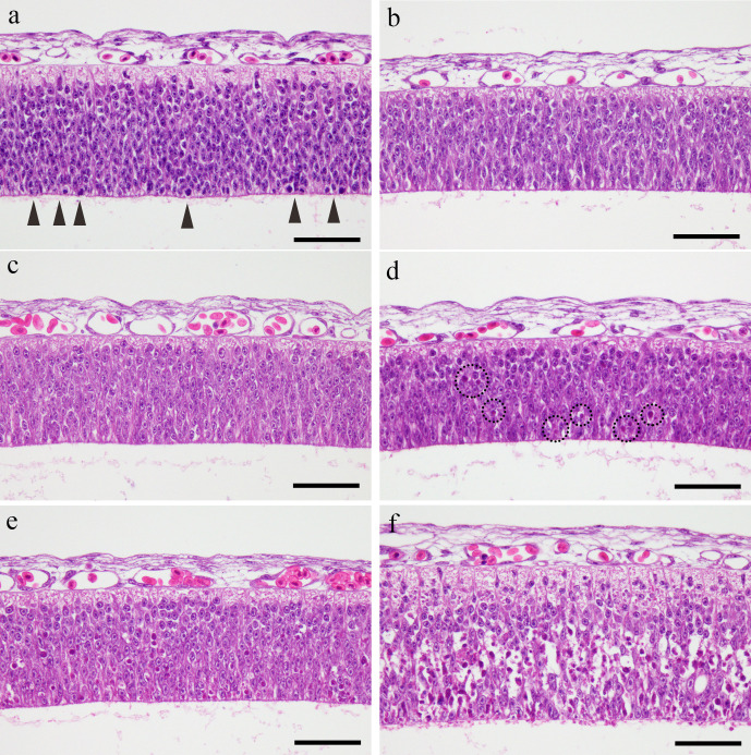
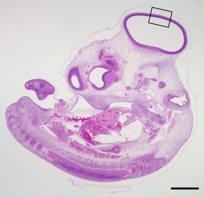
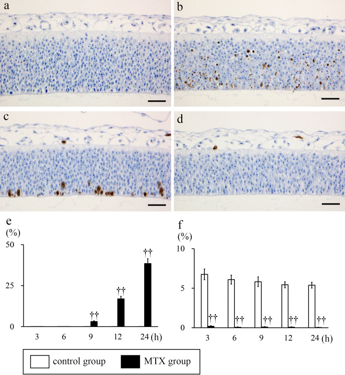
The optic tectum of Japanese quail embryos with in ovo exposure to methotrexate 100 ng/g egg on embryonic day 4 was examined from 3 to 24 hour after treatment. At 9 hour after methotrexate exposure, several apoptotic neuroepithelial cells appeared in the ventricular zone of the optic tectum; these increased in number and were diffusely distributed throughout all layers of the ventricular zone of the optic tectum at 12 hour. At 24 hour, neuroepithelial cells in the ventricular zone of the optic tectum were eliminated and showed sparse cell density. Throughout the experimental period, proliferation of neuroepithelial cells in the ventricular zone of the optic tectum of methotrexate-treated embryos was inhibited. These results suggest that neuroepithelial cells in the ventricular zone of the optic tectum in Japanese quail embryos can be affected by folic acid antimetabolites, methotrexate, at an early embryonic stage.
期刊介绍:
JTP is a scientific journal that publishes original studies in the field of toxicological pathology and in a wide variety of other related fields. The main scope of the journal is listed below.
Administrative Opinions of Policymakers and Regulatory Agencies
Adverse Events
Carcinogenesis
Data of A Predominantly Negative Nature
Drug-Induced Hematologic Toxicity
Embryological Pathology
High Throughput Pathology
Historical Data of Experimental Animals
Immunohistochemical Analysis
Molecular Pathology
Nomenclature of Lesions
Non-mammal Toxicity Study
Result or Lesion Induced by Chemicals of Which Names Hidden on Account of the Authors
Technology and Methodology Related to Toxicological Pathology
Tumor Pathology; Neoplasia and Hyperplasia
Ultrastructural Analysis
Use of Animal Models.