{"title":"CB6F1-Tg rasH2小鼠原发性附睾鳞状细胞癌。","authors":"Manabu Ikeda, Toshihisa Fujiwara, Junko Fujishima, Airo Yukawa, Kinji Kobayashi","doi":"10.1293/tox.2022-0047","DOIUrl":null,"url":null,"abstract":"<p><p>In a 26-week carcinogenicity study in rasH2-Tg mice, squamous cell carcinoma on the epididymis was observed in a male mouse in the positive control group treated with N-methyl-N-nitrosourea. A 29-week-old male rasH2-Tg mouse that was euthanized 21 weeks after the administration of N-methyl-N-nitrosourea had a white-grayish mass on the left caput epididymis. The mass was nodular and consisted of pleomorphic tumor cells forming alveolar, sheeted, and trabecular structures suggesting epithelial tumor growth. These cells presented a cobblestone-like arrangement and formed intercellular bridges. Keratinization was infrequently observed. Periodic acid-methenamine-silver staining revealed argyrophilic fibrous structures around the alveolar structure of the tumor cells. Immunohistochemically, the tumor cells were positive for cytokeratin AE1/AE3 and cytokeratin 14 and negative for cytokeratin 5, p63, uroplakin III, vimentin, desmin, and αSMA. These immunohistochemical results suggested the tumor cells originated from the epididymal ducts. Metastatic lesions were observed in the mesenteric, inguinal, and pancreaticoduodenal lymph nodes. Based on these results, this tumor was diagnosed to be a primary squamous cell carcinoma of the epididymis. This is the first report of primary squamous cell carcinoma of the epididymis in a rasH2-Tg mouse.</p>","PeriodicalId":17437,"journal":{"name":"Journal of Toxicologic Pathology","volume":"35 4","pages":"349-353"},"PeriodicalIF":0.9000,"publicationDate":"2022-10-01","publicationTypes":"Journal Article","fieldsOfStudy":null,"isOpenAccess":false,"openAccessPdf":"https://ftp.ncbi.nlm.nih.gov/pub/pmc/oa_pdf/4d/60/tox-35-349.PMC9647212.pdf","citationCount":"0","resultStr":"{\"title\":\"Primary epididymis squamous cell carcinoma in a CB6F1-Tg rasH2 mouse.\",\"authors\":\"Manabu Ikeda, Toshihisa Fujiwara, Junko Fujishima, Airo Yukawa, Kinji Kobayashi\",\"doi\":\"10.1293/tox.2022-0047\",\"DOIUrl\":null,\"url\":null,\"abstract\":\"<p><p>In a 26-week carcinogenicity study in rasH2-Tg mice, squamous cell carcinoma on the epididymis was observed in a male mouse in the positive control group treated with N-methyl-N-nitrosourea. A 29-week-old male rasH2-Tg mouse that was euthanized 21 weeks after the administration of N-methyl-N-nitrosourea had a white-grayish mass on the left caput epididymis. The mass was nodular and consisted of pleomorphic tumor cells forming alveolar, sheeted, and trabecular structures suggesting epithelial tumor growth. These cells presented a cobblestone-like arrangement and formed intercellular bridges. Keratinization was infrequently observed. Periodic acid-methenamine-silver staining revealed argyrophilic fibrous structures around the alveolar structure of the tumor cells. Immunohistochemically, the tumor cells were positive for cytokeratin AE1/AE3 and cytokeratin 14 and negative for cytokeratin 5, p63, uroplakin III, vimentin, desmin, and αSMA. These immunohistochemical results suggested the tumor cells originated from the epididymal ducts. Metastatic lesions were observed in the mesenteric, inguinal, and pancreaticoduodenal lymph nodes. Based on these results, this tumor was diagnosed to be a primary squamous cell carcinoma of the epididymis. This is the first report of primary squamous cell carcinoma of the epididymis in a rasH2-Tg mouse.</p>\",\"PeriodicalId\":17437,\"journal\":{\"name\":\"Journal of Toxicologic Pathology\",\"volume\":\"35 4\",\"pages\":\"349-353\"},\"PeriodicalIF\":0.9000,\"publicationDate\":\"2022-10-01\",\"publicationTypes\":\"Journal Article\",\"fieldsOfStudy\":null,\"isOpenAccess\":false,\"openAccessPdf\":\"https://ftp.ncbi.nlm.nih.gov/pub/pmc/oa_pdf/4d/60/tox-35-349.PMC9647212.pdf\",\"citationCount\":\"0\",\"resultStr\":null,\"platform\":\"Semanticscholar\",\"paperid\":null,\"PeriodicalName\":\"Journal of Toxicologic Pathology\",\"FirstCategoryId\":\"3\",\"ListUrlMain\":\"https://doi.org/10.1293/tox.2022-0047\",\"RegionNum\":4,\"RegionCategory\":\"医学\",\"ArticlePicture\":[],\"TitleCN\":null,\"AbstractTextCN\":null,\"PMCID\":null,\"EPubDate\":\"2022/6/30 0:00:00\",\"PubModel\":\"Epub\",\"JCR\":\"Q4\",\"JCRName\":\"PATHOLOGY\",\"Score\":null,\"Total\":0}","platform":"Semanticscholar","paperid":null,"PeriodicalName":"Journal of Toxicologic Pathology","FirstCategoryId":"3","ListUrlMain":"https://doi.org/10.1293/tox.2022-0047","RegionNum":4,"RegionCategory":"医学","ArticlePicture":[],"TitleCN":null,"AbstractTextCN":null,"PMCID":null,"EPubDate":"2022/6/30 0:00:00","PubModel":"Epub","JCR":"Q4","JCRName":"PATHOLOGY","Score":null,"Total":0}
Primary epididymis squamous cell carcinoma in a CB6F1-Tg rasH2 mouse.
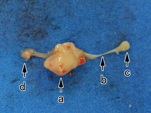
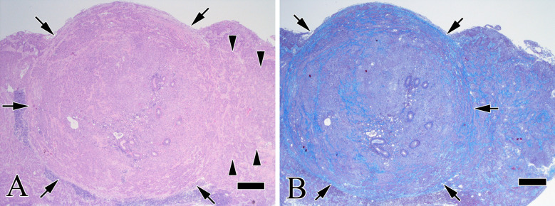
In a 26-week carcinogenicity study in rasH2-Tg mice, squamous cell carcinoma on the epididymis was observed in a male mouse in the positive control group treated with N-methyl-N-nitrosourea. A 29-week-old male rasH2-Tg mouse that was euthanized 21 weeks after the administration of N-methyl-N-nitrosourea had a white-grayish mass on the left caput epididymis. The mass was nodular and consisted of pleomorphic tumor cells forming alveolar, sheeted, and trabecular structures suggesting epithelial tumor growth. These cells presented a cobblestone-like arrangement and formed intercellular bridges. Keratinization was infrequently observed. Periodic acid-methenamine-silver staining revealed argyrophilic fibrous structures around the alveolar structure of the tumor cells. Immunohistochemically, the tumor cells were positive for cytokeratin AE1/AE3 and cytokeratin 14 and negative for cytokeratin 5, p63, uroplakin III, vimentin, desmin, and αSMA. These immunohistochemical results suggested the tumor cells originated from the epididymal ducts. Metastatic lesions were observed in the mesenteric, inguinal, and pancreaticoduodenal lymph nodes. Based on these results, this tumor was diagnosed to be a primary squamous cell carcinoma of the epididymis. This is the first report of primary squamous cell carcinoma of the epididymis in a rasH2-Tg mouse.
期刊介绍:
JTP is a scientific journal that publishes original studies in the field of toxicological pathology and in a wide variety of other related fields. The main scope of the journal is listed below.
Administrative Opinions of Policymakers and Regulatory Agencies
Adverse Events
Carcinogenesis
Data of A Predominantly Negative Nature
Drug-Induced Hematologic Toxicity
Embryological Pathology
High Throughput Pathology
Historical Data of Experimental Animals
Immunohistochemical Analysis
Molecular Pathology
Nomenclature of Lesions
Non-mammal Toxicity Study
Result or Lesion Induced by Chemicals of Which Names Hidden on Account of the Authors
Technology and Methodology Related to Toxicological Pathology
Tumor Pathology; Neoplasia and Hyperplasia
Ultrastructural Analysis
Use of Animal Models.

 求助内容:
求助内容: 应助结果提醒方式:


