{"title":"自动深度学习算法在胸片异常可靠筛查中的作用:一项前瞻性多中心质量改善研究。","authors":"Arunkumar Govindarajan, Aarthi Govindarajan, Swetha Tanamala, Subhankar Chattoraj, Bhargava Reddy, Rohitashva Agrawal, Divya Iyer, Anumeha Srivastava, Pradeep Kumar, Preetham Putha","doi":"10.3390/diagnostics12112724","DOIUrl":null,"url":null,"abstract":"<p><p>In medical practice, chest X-rays are the most ubiquitous diagnostic imaging tests. However, the current workload in extensive health care facilities and lack of well-trained radiologists is a significant challenge in the patient care pathway. Therefore, an accurate, reliable, and fast computer-aided diagnosis (CAD) system capable of detecting abnormalities in chest X-rays is crucial in improving the radiological workflow. In this prospective multicenter quality-improvement study, we have evaluated whether artificial intelligence (AI) can be used as a chest X-ray screening tool in real clinical settings. <b>Methods:</b> A team of radiologists used the AI-based chest X-ray screening tool (qXR) as a part of their daily reporting routine to report consecutive chest X-rays for this prospective multicentre study. This study took place in a large radiology network in India between June 2021 and March 2022. <b>Results:</b> A total of 65,604 chest X-rays were processed during the study period. The overall performance of AI achieved in detecting normal and abnormal chest X-rays was good. The high negatively predicted value (NPV) of 98.9% was achieved. The AI performance in terms of area under the curve (AUC), NPV for the corresponding subabnormalities obtained were blunted CP angle (0.97, 99.5%), hilar dysmorphism (0.86, 99.9%), cardiomegaly (0.96, 99.7%), reticulonodular pattern (0.91, 99.9%), rib fracture (0.98, 99.9%), scoliosis (0.98, 99.9%), atelectasis (0.96, 99.9%), calcification (0.96, 99.7%), consolidation (0.95, 99.6%), emphysema (0.96, 99.9%), fibrosis (0.95, 99.7%), nodule (0.91, 99.8%), opacity (0.92, 99.2%), pleural effusion (0.97, 99.7%), and pneumothorax (0.99, 99.9%). Additionally, the turnaround time (TAT) decreased by about 40.63% from pre-qXR period to post-qXR period. <b>Conclusions:</b> The AI-based chest X-ray solution (qXR) screened chest X-rays and assisted in ruling out normal patients with high confidence, thus allowing the radiologists to focus more on assessing pathology on abnormal chest X-rays and treatment pathways.</p>","PeriodicalId":520604,"journal":{"name":"Diagnostics (Basel, Switzerland)","volume":" ","pages":""},"PeriodicalIF":3.3000,"publicationDate":"2022-11-07","publicationTypes":"Journal Article","fieldsOfStudy":null,"isOpenAccess":false,"openAccessPdf":"https://www.ncbi.nlm.nih.gov/pmc/articles/PMC9689183/pdf/","citationCount":"4","resultStr":"{\"title\":\"Role of an Automated Deep Learning Algorithm for Reliable Screening of Abnormality in Chest Radiographs: A Prospective Multicenter Quality Improvement Study.\",\"authors\":\"Arunkumar Govindarajan, Aarthi Govindarajan, Swetha Tanamala, Subhankar Chattoraj, Bhargava Reddy, Rohitashva Agrawal, Divya Iyer, Anumeha Srivastava, Pradeep Kumar, Preetham Putha\",\"doi\":\"10.3390/diagnostics12112724\",\"DOIUrl\":null,\"url\":null,\"abstract\":\"<p><p>In medical practice, chest X-rays are the most ubiquitous diagnostic imaging tests. However, the current workload in extensive health care facilities and lack of well-trained radiologists is a significant challenge in the patient care pathway. Therefore, an accurate, reliable, and fast computer-aided diagnosis (CAD) system capable of detecting abnormalities in chest X-rays is crucial in improving the radiological workflow. In this prospective multicenter quality-improvement study, we have evaluated whether artificial intelligence (AI) can be used as a chest X-ray screening tool in real clinical settings. <b>Methods:</b> A team of radiologists used the AI-based chest X-ray screening tool (qXR) as a part of their daily reporting routine to report consecutive chest X-rays for this prospective multicentre study. This study took place in a large radiology network in India between June 2021 and March 2022. <b>Results:</b> A total of 65,604 chest X-rays were processed during the study period. The overall performance of AI achieved in detecting normal and abnormal chest X-rays was good. The high negatively predicted value (NPV) of 98.9% was achieved. The AI performance in terms of area under the curve (AUC), NPV for the corresponding subabnormalities obtained were blunted CP angle (0.97, 99.5%), hilar dysmorphism (0.86, 99.9%), cardiomegaly (0.96, 99.7%), reticulonodular pattern (0.91, 99.9%), rib fracture (0.98, 99.9%), scoliosis (0.98, 99.9%), atelectasis (0.96, 99.9%), calcification (0.96, 99.7%), consolidation (0.95, 99.6%), emphysema (0.96, 99.9%), fibrosis (0.95, 99.7%), nodule (0.91, 99.8%), opacity (0.92, 99.2%), pleural effusion (0.97, 99.7%), and pneumothorax (0.99, 99.9%). Additionally, the turnaround time (TAT) decreased by about 40.63% from pre-qXR period to post-qXR period. <b>Conclusions:</b> The AI-based chest X-ray solution (qXR) screened chest X-rays and assisted in ruling out normal patients with high confidence, thus allowing the radiologists to focus more on assessing pathology on abnormal chest X-rays and treatment pathways.</p>\",\"PeriodicalId\":520604,\"journal\":{\"name\":\"Diagnostics (Basel, Switzerland)\",\"volume\":\" \",\"pages\":\"\"},\"PeriodicalIF\":3.3000,\"publicationDate\":\"2022-11-07\",\"publicationTypes\":\"Journal Article\",\"fieldsOfStudy\":null,\"isOpenAccess\":false,\"openAccessPdf\":\"https://www.ncbi.nlm.nih.gov/pmc/articles/PMC9689183/pdf/\",\"citationCount\":\"4\",\"resultStr\":null,\"platform\":\"Semanticscholar\",\"paperid\":null,\"PeriodicalName\":\"Diagnostics (Basel, Switzerland)\",\"FirstCategoryId\":\"3\",\"ListUrlMain\":\"https://doi.org/10.3390/diagnostics12112724\",\"RegionNum\":0,\"RegionCategory\":null,\"ArticlePicture\":[],\"TitleCN\":null,\"AbstractTextCN\":null,\"PMCID\":null,\"EPubDate\":\"\",\"PubModel\":\"\",\"JCR\":\"\",\"JCRName\":\"\",\"Score\":null,\"Total\":0}","platform":"Semanticscholar","paperid":null,"PeriodicalName":"Diagnostics (Basel, Switzerland)","FirstCategoryId":"3","ListUrlMain":"https://doi.org/10.3390/diagnostics12112724","RegionNum":0,"RegionCategory":null,"ArticlePicture":[],"TitleCN":null,"AbstractTextCN":null,"PMCID":null,"EPubDate":"","PubModel":"","JCR":"","JCRName":"","Score":null,"Total":0}
Role of an Automated Deep Learning Algorithm for Reliable Screening of Abnormality in Chest Radiographs: A Prospective Multicenter Quality Improvement Study.
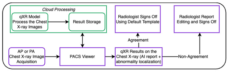
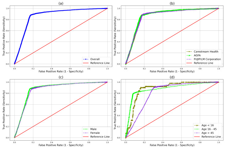
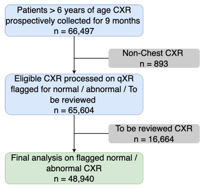
In medical practice, chest X-rays are the most ubiquitous diagnostic imaging tests. However, the current workload in extensive health care facilities and lack of well-trained radiologists is a significant challenge in the patient care pathway. Therefore, an accurate, reliable, and fast computer-aided diagnosis (CAD) system capable of detecting abnormalities in chest X-rays is crucial in improving the radiological workflow. In this prospective multicenter quality-improvement study, we have evaluated whether artificial intelligence (AI) can be used as a chest X-ray screening tool in real clinical settings. Methods: A team of radiologists used the AI-based chest X-ray screening tool (qXR) as a part of their daily reporting routine to report consecutive chest X-rays for this prospective multicentre study. This study took place in a large radiology network in India between June 2021 and March 2022. Results: A total of 65,604 chest X-rays were processed during the study period. The overall performance of AI achieved in detecting normal and abnormal chest X-rays was good. The high negatively predicted value (NPV) of 98.9% was achieved. The AI performance in terms of area under the curve (AUC), NPV for the corresponding subabnormalities obtained were blunted CP angle (0.97, 99.5%), hilar dysmorphism (0.86, 99.9%), cardiomegaly (0.96, 99.7%), reticulonodular pattern (0.91, 99.9%), rib fracture (0.98, 99.9%), scoliosis (0.98, 99.9%), atelectasis (0.96, 99.9%), calcification (0.96, 99.7%), consolidation (0.95, 99.6%), emphysema (0.96, 99.9%), fibrosis (0.95, 99.7%), nodule (0.91, 99.8%), opacity (0.92, 99.2%), pleural effusion (0.97, 99.7%), and pneumothorax (0.99, 99.9%). Additionally, the turnaround time (TAT) decreased by about 40.63% from pre-qXR period to post-qXR period. Conclusions: The AI-based chest X-ray solution (qXR) screened chest X-rays and assisted in ruling out normal patients with high confidence, thus allowing the radiologists to focus more on assessing pathology on abnormal chest X-rays and treatment pathways.

 求助内容:
求助内容: 应助结果提醒方式:


