{"title":"马胎儿期胃发育的组织学和组织计量学研究。","authors":"Dominik Poradowski, Aleksander Chrószcz","doi":"10.3390/ani12213047","DOIUrl":null,"url":null,"abstract":"<p><p>Histological and morphometrical analysis of the stomach wall was performed during the foetal period divided into three age groups (4th-11th month of gestation). The material was taken from non-glandular (the blind ventricular sac) and glandular parts (the plicated edge margin/cardiac part, the body of stomach and the pyloric part) of the stomach. It was preserved and prepared according to the standard protocol. The histological slides were stained (H-E, Masson-Goldner and PAS). The analyses were performed using the light microscope. All measurements were statistically elaborated. The crown-rump length growth rate was estimated as isometric. The blind ventricular sac growth rate was lower than CRL (negative allometric) and the partition of stomach mucosa into non-glandular and glandular part occurred in the 1st age group. The plicated edge margin/cardiac part and the pyloric part shoved similar tendencies. Only the body of stomach demonstrated a higher growth rate than CRL (positive allometric), which can be explained due to the strongest development of fundic glands. Moreover, comparing the adult reference group to the three parts of the foetal period, all metric values were lower than those achieved prenatally. The blind ventricular sac was covered with the multiple plane epithelium. The glandular parts of stomach that formed the superficial concave areas were covered with the simple columnar epithelium in the 1st age group, which developed to the cardiac, fundic, and pyloric glands in the 2rd and 3rd age groups. The <i>propria mucosae</i> was built with the mesenchyme, which differentiated later to the loose connective tissue. The muscular layer of mucosa was not clearly distinguishable in the 1st age group. The muscular layer of the stomach wall was formed with myoblasts in the 1st age group and later in the 2nd and the 3rd age groups built with fusiform myocytes divided into internal and external layers. The non-differentiated cells of glandular epithelium transformed into the parietal and chief cells. The first were visible in the gastric glands of the 2nd age group. Both of them were present in the 3rd age group gastric mucosa. The PAS staining proved a moderate PAS-positive reaction in the 2rd age group, while it was estimated as intense Pas-positive in the gastric glands in the 3rd age group and was comparable to postnatal observation (the adult reference group).</p>","PeriodicalId":519482,"journal":{"name":"Animals : an Open Access Journal from MDPI","volume":" ","pages":""},"PeriodicalIF":0.0000,"publicationDate":"2022-11-06","publicationTypes":"Journal Article","fieldsOfStudy":null,"isOpenAccess":false,"openAccessPdf":"https://www.ncbi.nlm.nih.gov/pmc/articles/PMC9656738/pdf/","citationCount":"1","resultStr":"{\"title\":\"Equine Stomach Development in the Foetal Period of Prenatal Life-A Histological and Histometric Study.\",\"authors\":\"Dominik Poradowski, Aleksander Chrószcz\",\"doi\":\"10.3390/ani12213047\",\"DOIUrl\":null,\"url\":null,\"abstract\":\"<p><p>Histological and morphometrical analysis of the stomach wall was performed during the foetal period divided into three age groups (4th-11th month of gestation). The material was taken from non-glandular (the blind ventricular sac) and glandular parts (the plicated edge margin/cardiac part, the body of stomach and the pyloric part) of the stomach. It was preserved and prepared according to the standard protocol. The histological slides were stained (H-E, Masson-Goldner and PAS). The analyses were performed using the light microscope. All measurements were statistically elaborated. The crown-rump length growth rate was estimated as isometric. The blind ventricular sac growth rate was lower than CRL (negative allometric) and the partition of stomach mucosa into non-glandular and glandular part occurred in the 1st age group. The plicated edge margin/cardiac part and the pyloric part shoved similar tendencies. Only the body of stomach demonstrated a higher growth rate than CRL (positive allometric), which can be explained due to the strongest development of fundic glands. Moreover, comparing the adult reference group to the three parts of the foetal period, all metric values were lower than those achieved prenatally. The blind ventricular sac was covered with the multiple plane epithelium. The glandular parts of stomach that formed the superficial concave areas were covered with the simple columnar epithelium in the 1st age group, which developed to the cardiac, fundic, and pyloric glands in the 2rd and 3rd age groups. The <i>propria mucosae</i> was built with the mesenchyme, which differentiated later to the loose connective tissue. The muscular layer of mucosa was not clearly distinguishable in the 1st age group. The muscular layer of the stomach wall was formed with myoblasts in the 1st age group and later in the 2nd and the 3rd age groups built with fusiform myocytes divided into internal and external layers. The non-differentiated cells of glandular epithelium transformed into the parietal and chief cells. The first were visible in the gastric glands of the 2nd age group. Both of them were present in the 3rd age group gastric mucosa. The PAS staining proved a moderate PAS-positive reaction in the 2rd age group, while it was estimated as intense Pas-positive in the gastric glands in the 3rd age group and was comparable to postnatal observation (the adult reference group).</p>\",\"PeriodicalId\":519482,\"journal\":{\"name\":\"Animals : an Open Access Journal from MDPI\",\"volume\":\" \",\"pages\":\"\"},\"PeriodicalIF\":0.0000,\"publicationDate\":\"2022-11-06\",\"publicationTypes\":\"Journal Article\",\"fieldsOfStudy\":null,\"isOpenAccess\":false,\"openAccessPdf\":\"https://www.ncbi.nlm.nih.gov/pmc/articles/PMC9656738/pdf/\",\"citationCount\":\"1\",\"resultStr\":null,\"platform\":\"Semanticscholar\",\"paperid\":null,\"PeriodicalName\":\"Animals : an Open Access Journal from MDPI\",\"FirstCategoryId\":\"97\",\"ListUrlMain\":\"https://doi.org/10.3390/ani12213047\",\"RegionNum\":0,\"RegionCategory\":null,\"ArticlePicture\":[],\"TitleCN\":null,\"AbstractTextCN\":null,\"PMCID\":null,\"EPubDate\":\"\",\"PubModel\":\"\",\"JCR\":\"\",\"JCRName\":\"\",\"Score\":null,\"Total\":0}","platform":"Semanticscholar","paperid":null,"PeriodicalName":"Animals : an Open Access Journal from MDPI","FirstCategoryId":"97","ListUrlMain":"https://doi.org/10.3390/ani12213047","RegionNum":0,"RegionCategory":null,"ArticlePicture":[],"TitleCN":null,"AbstractTextCN":null,"PMCID":null,"EPubDate":"","PubModel":"","JCR":"","JCRName":"","Score":null,"Total":0}
Equine Stomach Development in the Foetal Period of Prenatal Life-A Histological and Histometric Study.
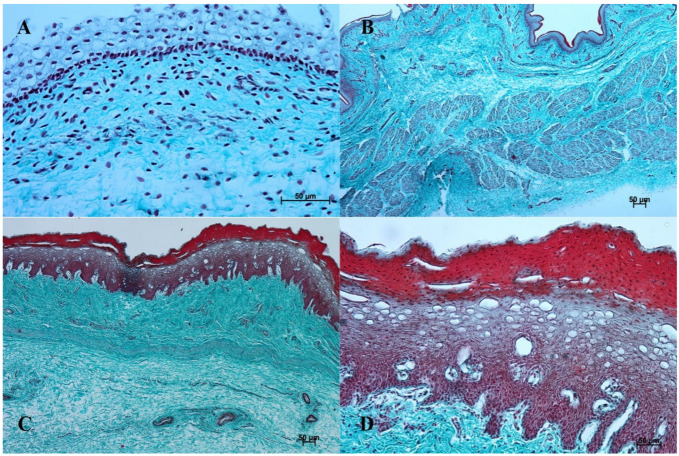
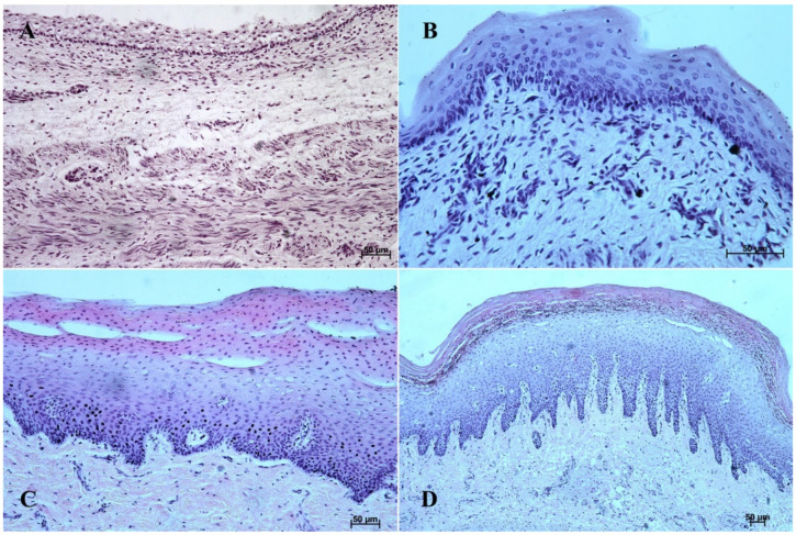
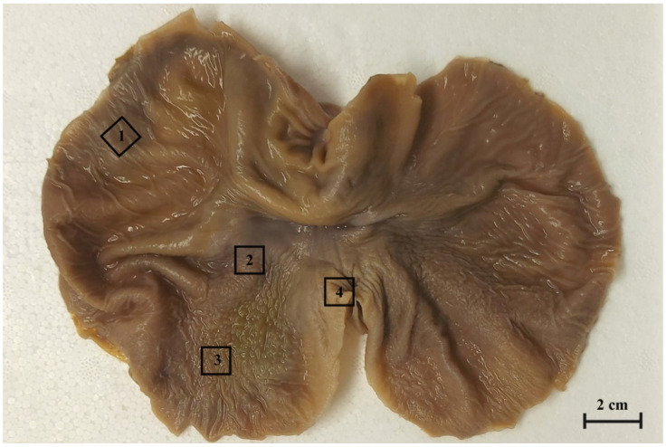
Histological and morphometrical analysis of the stomach wall was performed during the foetal period divided into three age groups (4th-11th month of gestation). The material was taken from non-glandular (the blind ventricular sac) and glandular parts (the plicated edge margin/cardiac part, the body of stomach and the pyloric part) of the stomach. It was preserved and prepared according to the standard protocol. The histological slides were stained (H-E, Masson-Goldner and PAS). The analyses were performed using the light microscope. All measurements were statistically elaborated. The crown-rump length growth rate was estimated as isometric. The blind ventricular sac growth rate was lower than CRL (negative allometric) and the partition of stomach mucosa into non-glandular and glandular part occurred in the 1st age group. The plicated edge margin/cardiac part and the pyloric part shoved similar tendencies. Only the body of stomach demonstrated a higher growth rate than CRL (positive allometric), which can be explained due to the strongest development of fundic glands. Moreover, comparing the adult reference group to the three parts of the foetal period, all metric values were lower than those achieved prenatally. The blind ventricular sac was covered with the multiple plane epithelium. The glandular parts of stomach that formed the superficial concave areas were covered with the simple columnar epithelium in the 1st age group, which developed to the cardiac, fundic, and pyloric glands in the 2rd and 3rd age groups. The propria mucosae was built with the mesenchyme, which differentiated later to the loose connective tissue. The muscular layer of mucosa was not clearly distinguishable in the 1st age group. The muscular layer of the stomach wall was formed with myoblasts in the 1st age group and later in the 2nd and the 3rd age groups built with fusiform myocytes divided into internal and external layers. The non-differentiated cells of glandular epithelium transformed into the parietal and chief cells. The first were visible in the gastric glands of the 2nd age group. Both of them were present in the 3rd age group gastric mucosa. The PAS staining proved a moderate PAS-positive reaction in the 2rd age group, while it was estimated as intense Pas-positive in the gastric glands in the 3rd age group and was comparable to postnatal observation (the adult reference group).

 求助内容:
求助内容: 应助结果提醒方式:


