{"title":"在牛津内侧单室膝关节置换术中,髓内棒的插入使股骨假体更外侧。","authors":"Toshikazu Tanaka, Yoshihito Suda, Tomoyuki Kamenaga, Akira Saito, Takaaki Fujishiro, Koji Okamoto, Takafumi Hiranaka","doi":"10.1186/s43019-022-00171-1","DOIUrl":null,"url":null,"abstract":"<p><strong>Background: </strong>This study aims to assess the influence of intramedullary rods on the implantation positions of femoral components using Microplasty instrumentation in Oxford unicompartmental knee arthroplasty. We hypothesized that femoral components can be laterally implanted incorrectly when using intramedullary rods.</p><p><strong>Methods: </strong>This prospective study included all 45 consecutive patients (53 knees) who underwent Oxford unicompartmental knee arthroplasty surgery for anteromedial osteoarthritis or spontaneous osteonecrosis of the knee at our hospital during the study period. A custom-made toolset comprising a triangular caliper and circular trial bearings was used to evaluate the distance between the bearing and the vertical wall of the tibia implant (wall-bearing space) using the caliper at 90° flexion both with and without intramedullary rods.</p><p><strong>Results: </strong>The wall-bearing space was significantly larger when the intramedullary rod was used than when intramedullary rod was not used (1.8 ± 1.1 mm versus 3.4 ± 1.2 mm, P < 0.001). The mean difference of wall-bearing space with and without intramedullary rod was 1.6 ± 0.7 mm.</p><p><strong>Conclusions: </strong>Femoral components can be laterally implanted incorrectly by an average of 1.6 mm when using intramedullary rods. The wall-bearing space should be evaluated using trial components, and if the relationship is improper, it should be corrected before keel slot preparation.</p>","PeriodicalId":17886,"journal":{"name":"Knee Surgery & Related Research","volume":" ","pages":"43"},"PeriodicalIF":4.4000,"publicationDate":"2022-11-11","publicationTypes":"Journal Article","fieldsOfStudy":null,"isOpenAccess":false,"openAccessPdf":"https://www.ncbi.nlm.nih.gov/pmc/articles/PMC9652838/pdf/","citationCount":"0","resultStr":"{\"title\":\"Intramedullary rod insertion places the femoral component more laterally during Oxford medial unicompartmental knee arthroplasty.\",\"authors\":\"Toshikazu Tanaka, Yoshihito Suda, Tomoyuki Kamenaga, Akira Saito, Takaaki Fujishiro, Koji Okamoto, Takafumi Hiranaka\",\"doi\":\"10.1186/s43019-022-00171-1\",\"DOIUrl\":null,\"url\":null,\"abstract\":\"<p><strong>Background: </strong>This study aims to assess the influence of intramedullary rods on the implantation positions of femoral components using Microplasty instrumentation in Oxford unicompartmental knee arthroplasty. We hypothesized that femoral components can be laterally implanted incorrectly when using intramedullary rods.</p><p><strong>Methods: </strong>This prospective study included all 45 consecutive patients (53 knees) who underwent Oxford unicompartmental knee arthroplasty surgery for anteromedial osteoarthritis or spontaneous osteonecrosis of the knee at our hospital during the study period. A custom-made toolset comprising a triangular caliper and circular trial bearings was used to evaluate the distance between the bearing and the vertical wall of the tibia implant (wall-bearing space) using the caliper at 90° flexion both with and without intramedullary rods.</p><p><strong>Results: </strong>The wall-bearing space was significantly larger when the intramedullary rod was used than when intramedullary rod was not used (1.8 ± 1.1 mm versus 3.4 ± 1.2 mm, P < 0.001). The mean difference of wall-bearing space with and without intramedullary rod was 1.6 ± 0.7 mm.</p><p><strong>Conclusions: </strong>Femoral components can be laterally implanted incorrectly by an average of 1.6 mm when using intramedullary rods. The wall-bearing space should be evaluated using trial components, and if the relationship is improper, it should be corrected before keel slot preparation.</p>\",\"PeriodicalId\":17886,\"journal\":{\"name\":\"Knee Surgery & Related Research\",\"volume\":\" \",\"pages\":\"43\"},\"PeriodicalIF\":4.4000,\"publicationDate\":\"2022-11-11\",\"publicationTypes\":\"Journal Article\",\"fieldsOfStudy\":null,\"isOpenAccess\":false,\"openAccessPdf\":\"https://www.ncbi.nlm.nih.gov/pmc/articles/PMC9652838/pdf/\",\"citationCount\":\"0\",\"resultStr\":null,\"platform\":\"Semanticscholar\",\"paperid\":null,\"PeriodicalName\":\"Knee Surgery & Related Research\",\"FirstCategoryId\":\"1085\",\"ListUrlMain\":\"https://doi.org/10.1186/s43019-022-00171-1\",\"RegionNum\":0,\"RegionCategory\":null,\"ArticlePicture\":[],\"TitleCN\":null,\"AbstractTextCN\":null,\"PMCID\":null,\"EPubDate\":\"\",\"PubModel\":\"\",\"JCR\":\"Q1\",\"JCRName\":\"ORTHOPEDICS\",\"Score\":null,\"Total\":0}","platform":"Semanticscholar","paperid":null,"PeriodicalName":"Knee Surgery & Related Research","FirstCategoryId":"1085","ListUrlMain":"https://doi.org/10.1186/s43019-022-00171-1","RegionNum":0,"RegionCategory":null,"ArticlePicture":[],"TitleCN":null,"AbstractTextCN":null,"PMCID":null,"EPubDate":"","PubModel":"","JCR":"Q1","JCRName":"ORTHOPEDICS","Score":null,"Total":0}
Intramedullary rod insertion places the femoral component more laterally during Oxford medial unicompartmental knee arthroplasty.
Background: This study aims to assess the influence of intramedullary rods on the implantation positions of femoral components using Microplasty instrumentation in Oxford unicompartmental knee arthroplasty. We hypothesized that femoral components can be laterally implanted incorrectly when using intramedullary rods.
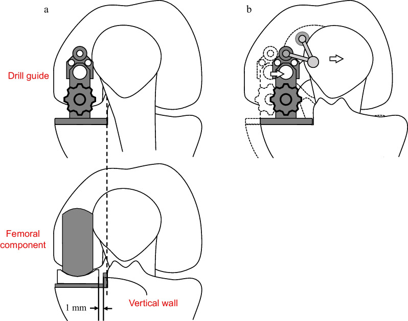
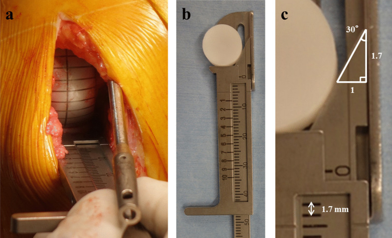
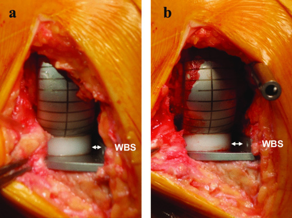
Methods: This prospective study included all 45 consecutive patients (53 knees) who underwent Oxford unicompartmental knee arthroplasty surgery for anteromedial osteoarthritis or spontaneous osteonecrosis of the knee at our hospital during the study period. A custom-made toolset comprising a triangular caliper and circular trial bearings was used to evaluate the distance between the bearing and the vertical wall of the tibia implant (wall-bearing space) using the caliper at 90° flexion both with and without intramedullary rods.
Results: The wall-bearing space was significantly larger when the intramedullary rod was used than when intramedullary rod was not used (1.8 ± 1.1 mm versus 3.4 ± 1.2 mm, P < 0.001). The mean difference of wall-bearing space with and without intramedullary rod was 1.6 ± 0.7 mm.
Conclusions: Femoral components can be laterally implanted incorrectly by an average of 1.6 mm when using intramedullary rods. The wall-bearing space should be evaluated using trial components, and if the relationship is improper, it should be corrected before keel slot preparation.

 求助内容:
求助内容: 应助结果提醒方式:


