M B Sturm, B P Joshi, S R Owens, E J Seibel, T D Wang
{"title":"区分不确定胆道狭窄的多重成像策略:一项体内研究","authors":"M B Sturm, B P Joshi, S R Owens, E J Seibel, T D Wang","doi":"10.47690/wjghe.2020.3303","DOIUrl":null,"url":null,"abstract":"<p><strong>Introduction: </strong>Indeterminant biliary strictures can be either malignant or benign. Biliary intraepithelial neoplasia (BilIN) is the precursor lesion to cholangiocarcinoma, a deadly bile duct cancer. Current diagnostic methods are limited by inadequate amounts of cells and tissues collected.</p><p><strong>Aim: </strong>We aim to demonstrate use of fluorescently-labeled peptides specific for EGFR, claudin-1, and ErbB2 to perform multiplexed imaging of biliary neoplasia.</p><p><strong>Methods: </strong>Formalin fixed and paraffin embedded specimens resected from human biliary strictures were sectioned. A gastrointestinal pathologist used standard criteria to score immunohistochemistry from biliary neoplasia and adjacent normal epithelium from the same specimen. Peptides specific for EGFR, claudin-1, and ErbB2 were fluorescently-labeled with FITC, Cy5, and IRDye800, respectively. The fluorophores were chosen to provide spectral separation to distinguish the individual targets. Immuno fluorescence images were collected using confocal microscopy.</p><p><strong>Results: </strong>Target expression was validated using immunohistochemistry. Staining was visualized on the surface of biliary duct epithelial cells and not in the stroma. Greater fluorescence intensity was observed for peptide binding to biliary neoplasia by comparison with normal. The mean ratio for neoplasia-to-normal was 1.4, 1.7, and 1.6, respectively, and the average intensities were significantly greater for neoplasia than normal for each peptide. Peptides and antibody binding co-localized with correlation of ρ=0.64, 0.51 and 0.62, respectively.</p><p><strong>Conclusions: </strong>A panel of fluorescently-labeled peptides can distinguish BilIN and cholangiocarcinoma from normal biliary epithelium, and may be used for multiplexed imaging of indeterminant biliary strictures.</p>","PeriodicalId":93828,"journal":{"name":"World journal of gastroenterology, hepatology and endoscopy","volume":"3 3","pages":""},"PeriodicalIF":0.0000,"publicationDate":"2020-01-01","publicationTypes":"Journal Article","fieldsOfStudy":null,"isOpenAccess":false,"openAccessPdf":"https://www.ncbi.nlm.nih.gov/pmc/articles/PMC9637386/pdf/","citationCount":"0","resultStr":"{\"title\":\"Multiplexed Imaging Strategy to Distinguish Indeterminant Biliary Strictures: An <i>Ex Vivo</i> Study.\",\"authors\":\"M B Sturm, B P Joshi, S R Owens, E J Seibel, T D Wang\",\"doi\":\"10.47690/wjghe.2020.3303\",\"DOIUrl\":null,\"url\":null,\"abstract\":\"<p><strong>Introduction: </strong>Indeterminant biliary strictures can be either malignant or benign. Biliary intraepithelial neoplasia (BilIN) is the precursor lesion to cholangiocarcinoma, a deadly bile duct cancer. Current diagnostic methods are limited by inadequate amounts of cells and tissues collected.</p><p><strong>Aim: </strong>We aim to demonstrate use of fluorescently-labeled peptides specific for EGFR, claudin-1, and ErbB2 to perform multiplexed imaging of biliary neoplasia.</p><p><strong>Methods: </strong>Formalin fixed and paraffin embedded specimens resected from human biliary strictures were sectioned. A gastrointestinal pathologist used standard criteria to score immunohistochemistry from biliary neoplasia and adjacent normal epithelium from the same specimen. Peptides specific for EGFR, claudin-1, and ErbB2 were fluorescently-labeled with FITC, Cy5, and IRDye800, respectively. The fluorophores were chosen to provide spectral separation to distinguish the individual targets. Immuno fluorescence images were collected using confocal microscopy.</p><p><strong>Results: </strong>Target expression was validated using immunohistochemistry. Staining was visualized on the surface of biliary duct epithelial cells and not in the stroma. Greater fluorescence intensity was observed for peptide binding to biliary neoplasia by comparison with normal. The mean ratio for neoplasia-to-normal was 1.4, 1.7, and 1.6, respectively, and the average intensities were significantly greater for neoplasia than normal for each peptide. Peptides and antibody binding co-localized with correlation of ρ=0.64, 0.51 and 0.62, respectively.</p><p><strong>Conclusions: </strong>A panel of fluorescently-labeled peptides can distinguish BilIN and cholangiocarcinoma from normal biliary epithelium, and may be used for multiplexed imaging of indeterminant biliary strictures.</p>\",\"PeriodicalId\":93828,\"journal\":{\"name\":\"World journal of gastroenterology, hepatology and endoscopy\",\"volume\":\"3 3\",\"pages\":\"\"},\"PeriodicalIF\":0.0000,\"publicationDate\":\"2020-01-01\",\"publicationTypes\":\"Journal Article\",\"fieldsOfStudy\":null,\"isOpenAccess\":false,\"openAccessPdf\":\"https://www.ncbi.nlm.nih.gov/pmc/articles/PMC9637386/pdf/\",\"citationCount\":\"0\",\"resultStr\":null,\"platform\":\"Semanticscholar\",\"paperid\":null,\"PeriodicalName\":\"World journal of gastroenterology, hepatology and endoscopy\",\"FirstCategoryId\":\"1085\",\"ListUrlMain\":\"https://doi.org/10.47690/wjghe.2020.3303\",\"RegionNum\":0,\"RegionCategory\":null,\"ArticlePicture\":[],\"TitleCN\":null,\"AbstractTextCN\":null,\"PMCID\":null,\"EPubDate\":\"2020/10/8 0:00:00\",\"PubModel\":\"Epub\",\"JCR\":\"\",\"JCRName\":\"\",\"Score\":null,\"Total\":0}","platform":"Semanticscholar","paperid":null,"PeriodicalName":"World journal of gastroenterology, hepatology and endoscopy","FirstCategoryId":"1085","ListUrlMain":"https://doi.org/10.47690/wjghe.2020.3303","RegionNum":0,"RegionCategory":null,"ArticlePicture":[],"TitleCN":null,"AbstractTextCN":null,"PMCID":null,"EPubDate":"2020/10/8 0:00:00","PubModel":"Epub","JCR":"","JCRName":"","Score":null,"Total":0}
Multiplexed Imaging Strategy to Distinguish Indeterminant Biliary Strictures: An Ex Vivo Study.
Introduction: Indeterminant biliary strictures can be either malignant or benign. Biliary intraepithelial neoplasia (BilIN) is the precursor lesion to cholangiocarcinoma, a deadly bile duct cancer. Current diagnostic methods are limited by inadequate amounts of cells and tissues collected.
Aim: We aim to demonstrate use of fluorescently-labeled peptides specific for EGFR, claudin-1, and ErbB2 to perform multiplexed imaging of biliary neoplasia.
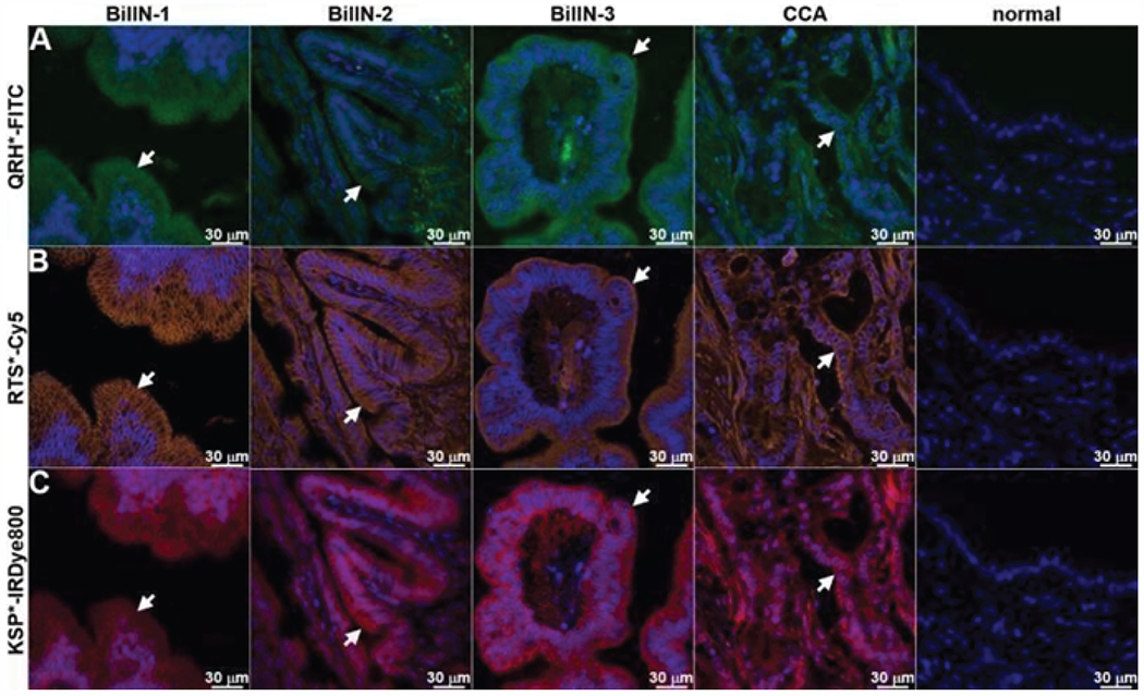
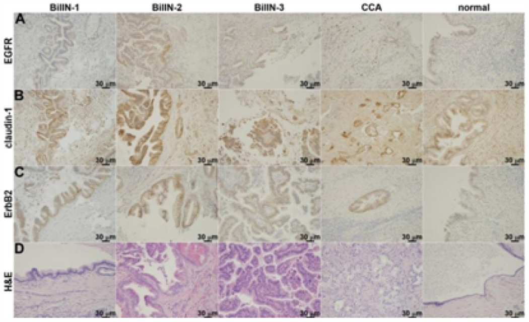
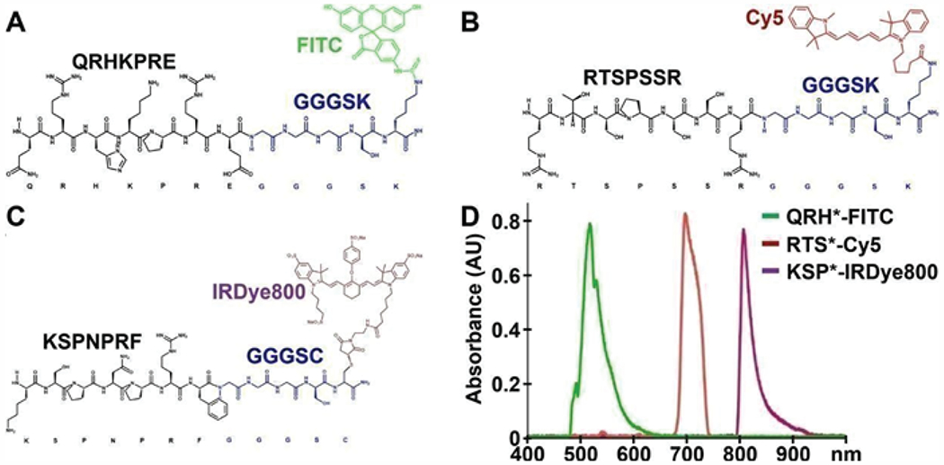
Methods: Formalin fixed and paraffin embedded specimens resected from human biliary strictures were sectioned. A gastrointestinal pathologist used standard criteria to score immunohistochemistry from biliary neoplasia and adjacent normal epithelium from the same specimen. Peptides specific for EGFR, claudin-1, and ErbB2 were fluorescently-labeled with FITC, Cy5, and IRDye800, respectively. The fluorophores were chosen to provide spectral separation to distinguish the individual targets. Immuno fluorescence images were collected using confocal microscopy.
Results: Target expression was validated using immunohistochemistry. Staining was visualized on the surface of biliary duct epithelial cells and not in the stroma. Greater fluorescence intensity was observed for peptide binding to biliary neoplasia by comparison with normal. The mean ratio for neoplasia-to-normal was 1.4, 1.7, and 1.6, respectively, and the average intensities were significantly greater for neoplasia than normal for each peptide. Peptides and antibody binding co-localized with correlation of ρ=0.64, 0.51 and 0.62, respectively.
Conclusions: A panel of fluorescently-labeled peptides can distinguish BilIN and cholangiocarcinoma from normal biliary epithelium, and may be used for multiplexed imaging of indeterminant biliary strictures.

 求助内容:
求助内容: 应助结果提醒方式:


