{"title":"一组斯里兰卡患者皮肤利什曼病的免疫病理学。","authors":"Harshima Disvini Wijesinghe, Gayani Kokila Wijesinghe, Deepika Fernando, Chandu de Silva","doi":"10.1177/2632010X221134804","DOIUrl":null,"url":null,"abstract":"<p><strong>Introduction and objectives: </strong><i>Leishmania donovani</i> is the causative organism of leishmaniasis in Sri Lanka. Studies on the immunopathology of leishmaniasis due to L. donovani are limited. The objective of this study was to describe the immunopathological characteristics of cutaneous leishmaniasis in a cohort of Sri Lankan patients.</p><p><strong>Methodology: </strong>Fifty skin biopsies of cutaneous leishmaniasis confirmed by detection of organisms by histology, culture, slit-skin smear, and/or polymerase chain reaction were reviewed. The inflammatory infiltrate was characterized by immunohistochemical staining for CD4, CD8, CD20, and CD68. Associations and correlations between immunohistochemical staining pattern and the parasitic load, and patterns of inflammation were determined.</p><p><strong>Results: </strong>The majority of biopsies showed a CD8+/CD4- T lymphocyte predominant infiltrate (84%, n = 42). A CD68 predominant infiltrate was seen in 16%(n = 8). The mean percentage of CD8+, CD4+, CD20+, and CD68+ inflammatory cells in the biopsies were 56.1% (SD = 16.5%), 2.6% (SD = 4.5%), 12.3% (SD = 10.9%), and 25.7% (SD = 15.8%) respectively. There was no association between the predominant inflammatory cell and the degree of inflammation (<i>P</i> = .173), presence of high RPI (<i>P</i> = .922), MRI(<i>P</i> = .367) or presence of granuloma (<i>P</i> = .247).The percentage of CD4+ cells showed a positive correlation with granuloma formation (Correlation coefficient = .411, <i>P</i> = .03). The percentage of CD20+ cells in the infiltrate showed a positive correlation with the degree of inflammation (Correlation coefficient = .491, <i>P</i> = .02) and the RPI (Correlation coefficient = .334, <i>P</i> = .018).</p><p><strong>Discussion and conclusion: </strong>Skin biopsies from cutaneous leishmaniasis due to <i>L. donovani</i> infection showed a CD8+/CD4- predominant infiltrate. This is similar to the findings of studies on cutaneous leishmaniasis due to some other species and suggests that the cytotoxic T cell response plays a role in infections due to <i>L. donovani</i>.</p>","PeriodicalId":53204,"journal":{"name":"Clinical Pathology","volume":" ","pages":"2632010X221134804"},"PeriodicalIF":1.9000,"publicationDate":"2022-11-02","publicationTypes":"Journal Article","fieldsOfStudy":null,"isOpenAccess":false,"openAccessPdf":"https://ftp.ncbi.nlm.nih.gov/pub/pmc/oa_pdf/27/86/10.1177_2632010X221134804.PMC9634189.pdf","citationCount":"1","resultStr":"{\"title\":\"Immunopathology of Cutaneous Leishmaniasis in a Cohort of Sri Lankan Patients.\",\"authors\":\"Harshima Disvini Wijesinghe, Gayani Kokila Wijesinghe, Deepika Fernando, Chandu de Silva\",\"doi\":\"10.1177/2632010X221134804\",\"DOIUrl\":null,\"url\":null,\"abstract\":\"<p><strong>Introduction and objectives: </strong><i>Leishmania donovani</i> is the causative organism of leishmaniasis in Sri Lanka. Studies on the immunopathology of leishmaniasis due to L. donovani are limited. The objective of this study was to describe the immunopathological characteristics of cutaneous leishmaniasis in a cohort of Sri Lankan patients.</p><p><strong>Methodology: </strong>Fifty skin biopsies of cutaneous leishmaniasis confirmed by detection of organisms by histology, culture, slit-skin smear, and/or polymerase chain reaction were reviewed. The inflammatory infiltrate was characterized by immunohistochemical staining for CD4, CD8, CD20, and CD68. Associations and correlations between immunohistochemical staining pattern and the parasitic load, and patterns of inflammation were determined.</p><p><strong>Results: </strong>The majority of biopsies showed a CD8+/CD4- T lymphocyte predominant infiltrate (84%, n = 42). A CD68 predominant infiltrate was seen in 16%(n = 8). The mean percentage of CD8+, CD4+, CD20+, and CD68+ inflammatory cells in the biopsies were 56.1% (SD = 16.5%), 2.6% (SD = 4.5%), 12.3% (SD = 10.9%), and 25.7% (SD = 15.8%) respectively. There was no association between the predominant inflammatory cell and the degree of inflammation (<i>P</i> = .173), presence of high RPI (<i>P</i> = .922), MRI(<i>P</i> = .367) or presence of granuloma (<i>P</i> = .247).The percentage of CD4+ cells showed a positive correlation with granuloma formation (Correlation coefficient = .411, <i>P</i> = .03). The percentage of CD20+ cells in the infiltrate showed a positive correlation with the degree of inflammation (Correlation coefficient = .491, <i>P</i> = .02) and the RPI (Correlation coefficient = .334, <i>P</i> = .018).</p><p><strong>Discussion and conclusion: </strong>Skin biopsies from cutaneous leishmaniasis due to <i>L. donovani</i> infection showed a CD8+/CD4- predominant infiltrate. This is similar to the findings of studies on cutaneous leishmaniasis due to some other species and suggests that the cytotoxic T cell response plays a role in infections due to <i>L. donovani</i>.</p>\",\"PeriodicalId\":53204,\"journal\":{\"name\":\"Clinical Pathology\",\"volume\":\" \",\"pages\":\"2632010X221134804\"},\"PeriodicalIF\":1.9000,\"publicationDate\":\"2022-11-02\",\"publicationTypes\":\"Journal Article\",\"fieldsOfStudy\":null,\"isOpenAccess\":false,\"openAccessPdf\":\"https://ftp.ncbi.nlm.nih.gov/pub/pmc/oa_pdf/27/86/10.1177_2632010X221134804.PMC9634189.pdf\",\"citationCount\":\"1\",\"resultStr\":null,\"platform\":\"Semanticscholar\",\"paperid\":null,\"PeriodicalName\":\"Clinical Pathology\",\"FirstCategoryId\":\"1085\",\"ListUrlMain\":\"https://doi.org/10.1177/2632010X221134804\",\"RegionNum\":0,\"RegionCategory\":null,\"ArticlePicture\":[],\"TitleCN\":null,\"AbstractTextCN\":null,\"PMCID\":null,\"EPubDate\":\"2022/1/1 0:00:00\",\"PubModel\":\"eCollection\",\"JCR\":\"Q3\",\"JCRName\":\"PATHOLOGY\",\"Score\":null,\"Total\":0}","platform":"Semanticscholar","paperid":null,"PeriodicalName":"Clinical Pathology","FirstCategoryId":"1085","ListUrlMain":"https://doi.org/10.1177/2632010X221134804","RegionNum":0,"RegionCategory":null,"ArticlePicture":[],"TitleCN":null,"AbstractTextCN":null,"PMCID":null,"EPubDate":"2022/1/1 0:00:00","PubModel":"eCollection","JCR":"Q3","JCRName":"PATHOLOGY","Score":null,"Total":0}
Immunopathology of Cutaneous Leishmaniasis in a Cohort of Sri Lankan Patients.
Introduction and objectives: Leishmania donovani is the causative organism of leishmaniasis in Sri Lanka. Studies on the immunopathology of leishmaniasis due to L. donovani are limited. The objective of this study was to describe the immunopathological characteristics of cutaneous leishmaniasis in a cohort of Sri Lankan patients.
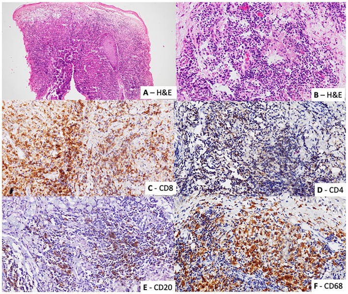
Methodology: Fifty skin biopsies of cutaneous leishmaniasis confirmed by detection of organisms by histology, culture, slit-skin smear, and/or polymerase chain reaction were reviewed. The inflammatory infiltrate was characterized by immunohistochemical staining for CD4, CD8, CD20, and CD68. Associations and correlations between immunohistochemical staining pattern and the parasitic load, and patterns of inflammation were determined.
Results: The majority of biopsies showed a CD8+/CD4- T lymphocyte predominant infiltrate (84%, n = 42). A CD68 predominant infiltrate was seen in 16%(n = 8). The mean percentage of CD8+, CD4+, CD20+, and CD68+ inflammatory cells in the biopsies were 56.1% (SD = 16.5%), 2.6% (SD = 4.5%), 12.3% (SD = 10.9%), and 25.7% (SD = 15.8%) respectively. There was no association between the predominant inflammatory cell and the degree of inflammation (P = .173), presence of high RPI (P = .922), MRI(P = .367) or presence of granuloma (P = .247).The percentage of CD4+ cells showed a positive correlation with granuloma formation (Correlation coefficient = .411, P = .03). The percentage of CD20+ cells in the infiltrate showed a positive correlation with the degree of inflammation (Correlation coefficient = .491, P = .02) and the RPI (Correlation coefficient = .334, P = .018).
Discussion and conclusion: Skin biopsies from cutaneous leishmaniasis due to L. donovani infection showed a CD8+/CD4- predominant infiltrate. This is similar to the findings of studies on cutaneous leishmaniasis due to some other species and suggests that the cytotoxic T cell response plays a role in infections due to L. donovani.

 求助内容:
求助内容: 应助结果提醒方式:


