Marianne Flinck, Johan von Heideken, Ylva Aurell, Jacques Riad
{"title":"弹性髓内钉治疗儿童股骨干骨折后骨骼成熟后的腿长差异。","authors":"Marianne Flinck, Johan von Heideken, Ylva Aurell, Jacques Riad","doi":"10.1177/18632521221106388","DOIUrl":null,"url":null,"abstract":"<p><strong>Purpose: </strong>The purpose was to study radiographic and perceived leg length discrepancy after skeletal maturity in patients treated for femoral shaft fractures with elastic stable intramedullary nails in childhood.</p><p><strong>Methods: </strong>Thirty-five adults underwent standing radiographs and answered a questionnaire regarding perception of leg length discrepancy. Demographic data, fracture characteristics, angulation, stability of fixation, and callus formation, at time of fracture, were assessed.</p><p><strong>Results: </strong>Mean age at fracture was 10.2 (4.9-16.7) years, and mean follow-up time was 11.1 (3.8-16.8) years. In 8 of 35 participants, the fractured limb was 11-15 mm longer than the non-fractured, and in 16, 1-10 mm longer. In eight participants, the fractured limb was 1-10 mm shorter than the non-fractured, and in three participants, 12-23 mm shorter. The younger the child, the greater the lengthening (R<sub>s</sub> = -0.49, p = 0.003). The greater the femoral angulation at time of fracture, the greater the shortening (R<sub>s</sub> = 0.42, p = 0.013). There was no significant correlation between stability of fixation or callus formation 1 month postoperatively and radiographic leg length discrepancy after skeletal maturity. Fourteen (40%) had perception of leg length discrepancy at follow-up, of whom eight had a radiographic leg length discrepancy of 10-24 mm.</p><p><strong>Conclusion: </strong>Treatment with elastic stable intramedullary nail of femoral shaft fracture in childhood may result in radiographic leg length discrepancy. Younger children were more prone to lengthening and should possibly be assessed before skeletal maturity. The degree of fracture stability or callus formation at the time of fracture did not significantly affect leg length discrepancy. Perception of leg length discrepancy was not necessarily associated with a radiographic leg length discrepancy (≥10 mm).</p><p><strong>Level of evidence: </strong>level IV, case series.</p>","PeriodicalId":138259,"journal":{"name":"Journal of Children's Orthopaedics","volume":" ","pages":"276-284"},"PeriodicalIF":0.0000,"publicationDate":"2022-08-01","publicationTypes":"Journal Article","fieldsOfStudy":null,"isOpenAccess":false,"openAccessPdf":"https://ftp.ncbi.nlm.nih.gov/pub/pmc/oa_pdf/1c/79/10.1177_18632521221106388.PMC9382705.pdf","citationCount":"2","resultStr":"{\"title\":\"Leg length discrepancy after skeletal maturity in patients treated with elastic intramedullary nails after femoral shaft fractures in childhood.\",\"authors\":\"Marianne Flinck, Johan von Heideken, Ylva Aurell, Jacques Riad\",\"doi\":\"10.1177/18632521221106388\",\"DOIUrl\":null,\"url\":null,\"abstract\":\"<p><strong>Purpose: </strong>The purpose was to study radiographic and perceived leg length discrepancy after skeletal maturity in patients treated for femoral shaft fractures with elastic stable intramedullary nails in childhood.</p><p><strong>Methods: </strong>Thirty-five adults underwent standing radiographs and answered a questionnaire regarding perception of leg length discrepancy. Demographic data, fracture characteristics, angulation, stability of fixation, and callus formation, at time of fracture, were assessed.</p><p><strong>Results: </strong>Mean age at fracture was 10.2 (4.9-16.7) years, and mean follow-up time was 11.1 (3.8-16.8) years. In 8 of 35 participants, the fractured limb was 11-15 mm longer than the non-fractured, and in 16, 1-10 mm longer. In eight participants, the fractured limb was 1-10 mm shorter than the non-fractured, and in three participants, 12-23 mm shorter. The younger the child, the greater the lengthening (R<sub>s</sub> = -0.49, p = 0.003). The greater the femoral angulation at time of fracture, the greater the shortening (R<sub>s</sub> = 0.42, p = 0.013). There was no significant correlation between stability of fixation or callus formation 1 month postoperatively and radiographic leg length discrepancy after skeletal maturity. Fourteen (40%) had perception of leg length discrepancy at follow-up, of whom eight had a radiographic leg length discrepancy of 10-24 mm.</p><p><strong>Conclusion: </strong>Treatment with elastic stable intramedullary nail of femoral shaft fracture in childhood may result in radiographic leg length discrepancy. Younger children were more prone to lengthening and should possibly be assessed before skeletal maturity. The degree of fracture stability or callus formation at the time of fracture did not significantly affect leg length discrepancy. Perception of leg length discrepancy was not necessarily associated with a radiographic leg length discrepancy (≥10 mm).</p><p><strong>Level of evidence: </strong>level IV, case series.</p>\",\"PeriodicalId\":138259,\"journal\":{\"name\":\"Journal of Children's Orthopaedics\",\"volume\":\" \",\"pages\":\"276-284\"},\"PeriodicalIF\":0.0000,\"publicationDate\":\"2022-08-01\",\"publicationTypes\":\"Journal Article\",\"fieldsOfStudy\":null,\"isOpenAccess\":false,\"openAccessPdf\":\"https://ftp.ncbi.nlm.nih.gov/pub/pmc/oa_pdf/1c/79/10.1177_18632521221106388.PMC9382705.pdf\",\"citationCount\":\"2\",\"resultStr\":null,\"platform\":\"Semanticscholar\",\"paperid\":null,\"PeriodicalName\":\"Journal of Children's Orthopaedics\",\"FirstCategoryId\":\"3\",\"ListUrlMain\":\"https://doi.org/10.1177/18632521221106388\",\"RegionNum\":0,\"RegionCategory\":null,\"ArticlePicture\":[],\"TitleCN\":null,\"AbstractTextCN\":null,\"PMCID\":null,\"EPubDate\":\"2022/8/2 0:00:00\",\"PubModel\":\"Epub\",\"JCR\":\"\",\"JCRName\":\"\",\"Score\":null,\"Total\":0}","platform":"Semanticscholar","paperid":null,"PeriodicalName":"Journal of Children's Orthopaedics","FirstCategoryId":"3","ListUrlMain":"https://doi.org/10.1177/18632521221106388","RegionNum":0,"RegionCategory":null,"ArticlePicture":[],"TitleCN":null,"AbstractTextCN":null,"PMCID":null,"EPubDate":"2022/8/2 0:00:00","PubModel":"Epub","JCR":"","JCRName":"","Score":null,"Total":0}
引用次数: 2
摘要
目的:研究儿童弹性稳定髓内钉治疗股骨干骨折患者骨骼成熟后的x线片和感知腿长差异。方法:35名成人接受站立x线摄影,并回答一份关于腿长差异感知的问卷。评估骨折时的人口学数据、骨折特征、角度、固定稳定性和骨痂形成。结果:骨折患者平均年龄10.2(4.9 ~ 16.7)岁,平均随访时间11.1(3.8 ~ 16.8)年。在35名参与者中,有8名骨折的肢体比未骨折的肢体长11-15毫米,有16名骨折的肢体长1-10毫米。在8名参与者中,骨折的肢体比未骨折的肢体短1-10毫米,在3名参与者中,短12-23毫米。儿童年龄越小,延长时间越长(Rs = -0.49, p = 0.003)。骨折时股骨成角越大,短缩越大(Rs = 0.42, p = 0.013)。术后1个月的固定物稳定性或骨痂形成与骨骼成熟后的x线腿长差异无显著相关性。14人(40%)在随访时感觉腿长差异,其中8人的x线片腿长差异为10-24毫米。结论:弹性稳定髓内钉治疗儿童股骨干骨折可导致x线片上的腿长差异。年龄较小的儿童更倾向于延长,应该在骨骼成熟之前进行评估。骨折时骨折稳定性或骨痂形成程度对腿长差异无显著影响。腿长差异的感知与x线片腿长差异(≥10 mm)并不一定相关。证据等级:四级,案例系列。
Leg length discrepancy after skeletal maturity in patients treated with elastic intramedullary nails after femoral shaft fractures in childhood.
Purpose: The purpose was to study radiographic and perceived leg length discrepancy after skeletal maturity in patients treated for femoral shaft fractures with elastic stable intramedullary nails in childhood.
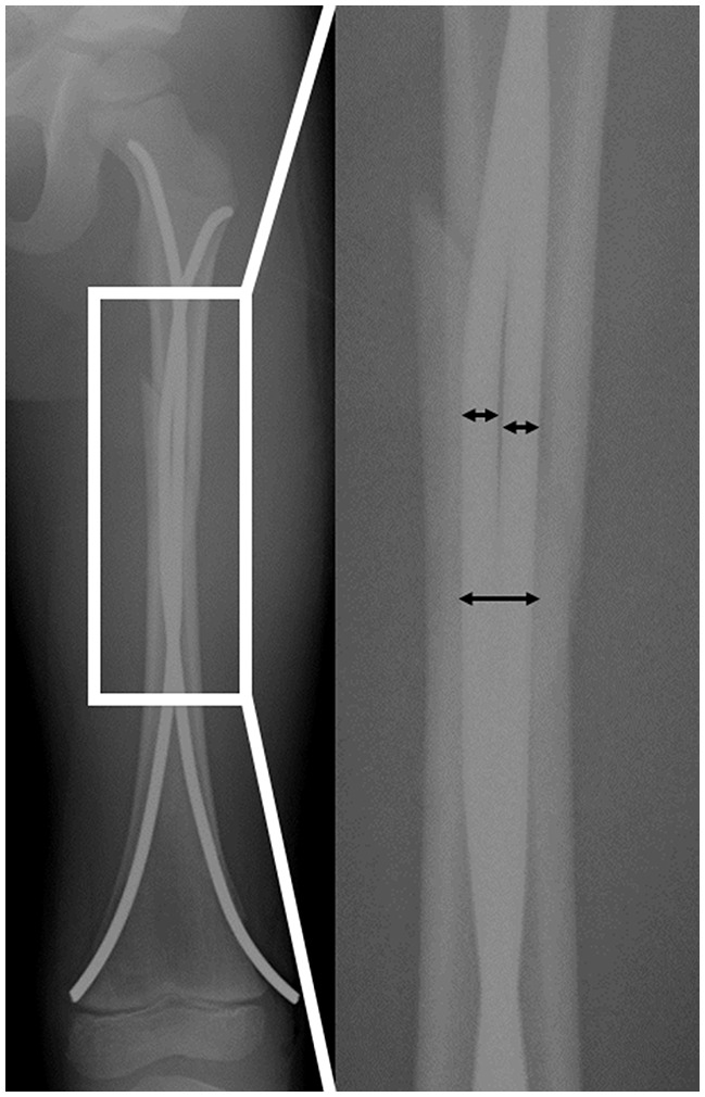
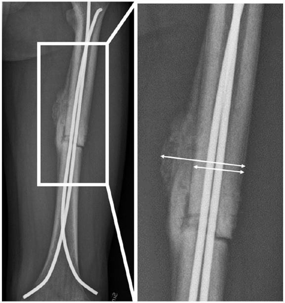
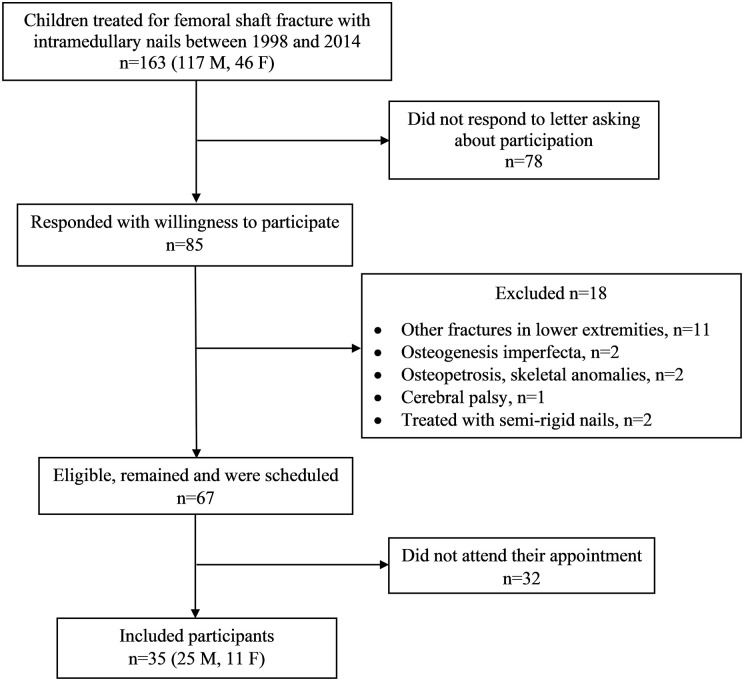
Methods: Thirty-five adults underwent standing radiographs and answered a questionnaire regarding perception of leg length discrepancy. Demographic data, fracture characteristics, angulation, stability of fixation, and callus formation, at time of fracture, were assessed.
Results: Mean age at fracture was 10.2 (4.9-16.7) years, and mean follow-up time was 11.1 (3.8-16.8) years. In 8 of 35 participants, the fractured limb was 11-15 mm longer than the non-fractured, and in 16, 1-10 mm longer. In eight participants, the fractured limb was 1-10 mm shorter than the non-fractured, and in three participants, 12-23 mm shorter. The younger the child, the greater the lengthening (Rs = -0.49, p = 0.003). The greater the femoral angulation at time of fracture, the greater the shortening (Rs = 0.42, p = 0.013). There was no significant correlation between stability of fixation or callus formation 1 month postoperatively and radiographic leg length discrepancy after skeletal maturity. Fourteen (40%) had perception of leg length discrepancy at follow-up, of whom eight had a radiographic leg length discrepancy of 10-24 mm.
Conclusion: Treatment with elastic stable intramedullary nail of femoral shaft fracture in childhood may result in radiographic leg length discrepancy. Younger children were more prone to lengthening and should possibly be assessed before skeletal maturity. The degree of fracture stability or callus formation at the time of fracture did not significantly affect leg length discrepancy. Perception of leg length discrepancy was not necessarily associated with a radiographic leg length discrepancy (≥10 mm).

 求助内容:
求助内容: 应助结果提醒方式:


